 WhatsApp

Subscribe to our
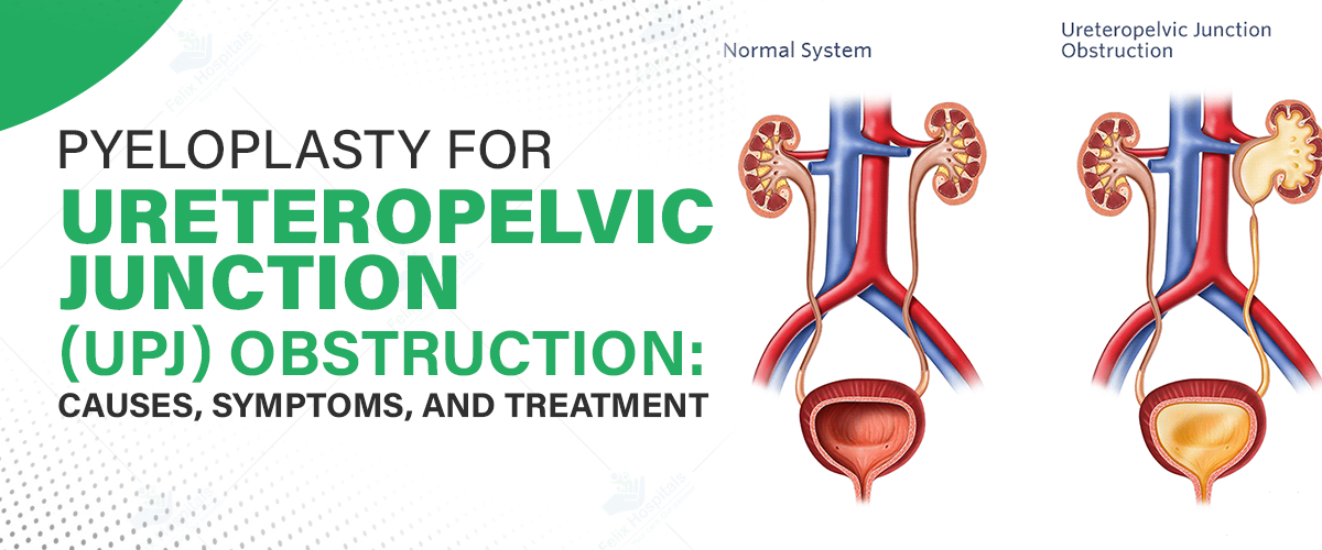
Ureteropelvic Junction (UPJ) Obstruction is a medical condition where there is a blockage at the junction where the renal pelvis meets the ureter. This obstruction can impede the normal flow of urine from the kidney to the bladder, leading to various complications, including kidney damage. Prompt diagnosis and treatment are crucial to prevent long-term kidney issues. If left untreated, UPJ obstruction can result in severe kidney damage, hypertension, and recurrent infections.
At the Top Pyeloplasty Surgery Hospital, specialists offer advanced treatments, with Pyeloplasty being the primary surgical solution to relieve the obstruction and restore proper kidney function. This procedure is highly effective, offering long-term relief and improving the patient’s quality of life.
If you're experiencing persistent urinary issues, seeking medical advice is crucial. Contact us today at +91 9667064100.
UPJ Obstruction occurs when there is a blockage at the point where the renal pelvis, the funnel-shaped part of the kidney, connects to the ureter. This blockage disrupts the flow of urine from the kidney to the bladder. Over time, this can lead to kidney swelling (hydronephrosis) and impairment of kidney function.
The condition can be congenital (present at birth) or acquired later in life due to various factors. Whether congenital or acquired, timely detection and intervention are necessary to avoid complications like kidney failure.
There are two primary categories of causes for UPJ Obstruction: congenital and acquired.
The symptoms of UPJ obstruction can vary depending on the severity and duration of the condition.
To diagnose UPJ Obstruction, several imaging tests and functional studies are performed.
Pyeloplasty is the surgical procedure used to treat UPJ Obstruction. The goal is to remove the obstruction and restore normal urine flow from the kidney to the bladder. Pyeloplasty has a high success rate and is the preferred method of treatment for UPJ obstruction, especially when conservative treatments are ineffective.
Types of Pyeloplasty
Preoperative Preparation
Surgical Process
Postoperative Care and Recovery
Like any surgery, Pyeloplasty carries potential risks, including:
For effective treatment of UPJ Obstruction, schedule a consultation with Dr. Bhanwar Lal Barkesia, one of the Best Pyeloplasty Doctors in Noida at Felix Hospitals. With years of expertise in urology, Dr. Barkesia offers personalized care and advanced surgical techniques to treat UPJ Obstruction and other urological issues.
Schedule an Appointment at Felix Hospitals for the best urology care and treatment options for UPJ Obstruction.
Early detection and timely treatment of UPJ Obstruction are essential to prevent further kidney damage and improve the patient’s quality of life. Thanks to advancements in minimally invasive surgical techniques like laparoscopic and robotic-assisted pyeloplasty, patients can expect a quicker recovery and fewer complications. When considering pyeloplasty, it is important to understand the pyeloplasty surgery cost in Noida and consult with experienced urologists to determine the best course of action for optimal health.
Q1- What are the long-term effects of untreated UPJ Obstruction on kidney function?
ANS: UPJ Obstruction can lead to chronic kidney damage, progressive loss of kidney function, and, in severe cases, kidney failure. Early intervention is essential to prevent these complications and preserve kidney health.
Q2- How can I differentiate between flank pain caused by UPJ Obstruction and other kidney-related conditions?
ANS: Flank pain due to UPJ Obstruction is often accompanied by symptoms like nausea, vomiting, and recurrent urinary tract infections (UTIs). Imaging tests such as ultrasound or CT scan can help confirm the diagnosis and rule out other kidney conditions.
Q3- What factors determine whether a patient should undergo laparoscopic, robotic-assisted, or open pyeloplasty?
ANS: The choice of surgical method depends on the severity of the obstruction, the patient’s overall health, and the experience of the surgeon. Robotic-assisted and laparoscopic procedures are generally preferred for their minimally invasive nature, offering quicker recovery and less post-surgical pain.
Q4- How does a renal scan help in diagnosing UPJ Obstruction, and what does it reveal?
ANS: A renal scan, also known as a diuretic renal scan, assesses how well the kidneys are draining urine. It helps evaluate the extent of the obstruction and the functionality of the affected kidney, providing essential information for treatment planning.
Q5- Is there a risk of UPJ Obstruction recurring after pyeloplasty, and how can this be prevented?
ANS: Although recurrence is rare, it can happen, especially if the original obstruction is not fully removed. Regular follow-up visits and imaging tests after surgery help monitor kidney function and detect any early signs of recurrence.
Q6- Can UPJ Obstruction be diagnosed during routine check-ups, or is it detected only when symptoms become severe?
ANS: While UPJ Obstruction may be asymptomatic in the early stages, it is often diagnosed during routine imaging tests for other conditions, such as UTIs or unexplained flank pain. Early detection through these tests can prevent further complications.
Q7- What dietary changes should be made after pyeloplasty to support kidney health?
ANS: After pyeloplasty, maintaining a kidney-friendly diet is essential. Patients are advised to stay hydrated, reduce salt intake, and eat a balanced diet rich in fruits, vegetables, and lean proteins to support optimal kidney recovery and long-term health.

Urological health is vital for well-being, influenced by diet and exercise. Good nutrition and activity help prevent conditions like UTIs, kidney stones, and cancers.
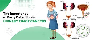
For instance, bladder cancer has a five-year survival rate of about 77% when diagnosed early, compared to just 5% in advanced stages. Early diagnosis directly correlates with improved outcomes and survival rates.
One of the best hospitals for urinary tract treatment in Noida offers comprehensive urology care, featuring expert urologists and advanced diagnostic and treatment facilities.

Our urology and oncology specialists bring extensive experience and expertise in treating various forms of urological cancer.

Subscribe to our
© Copyright 2026. All Rights Reserved by Felix Healthcare Private Limited
Design and development by :


 +(91)9667064100
 Emergency : +(91)9667064100