 WhatsApp

Subscribe to our
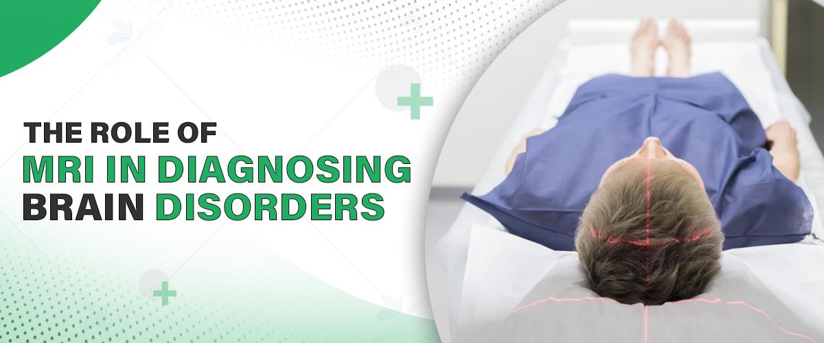
In terms of detecting neurological disorders, Magnetic Resonance Imaging (MRI) of the brain is perhaps the most sophisticated and utilized technology in use today. Whether the doctor is checking for recurring headaches, seizures, stroke-like symptoms, or other neurological issues, a brain MRI offers precise information that no other imaging technique can.
If you are in need of care or consultation, it is essential to seek out the best neurology hospital in Noida—a facility with state-of-the-art MRI technology and highly trained experts who can properly analyze your scans and direct treatment.
This in-depth guide will familiarize you with what a brain MRI is, how it is performed, what happens during the process and why your neurologist would prescribe this vital diagnostic test.
Early and precise diagnosis is the key to successful treatment. Don't hesitate—seek experts by calling +91 9667064100.
MRI of the brain is a painless imaging examination that employs a combination of powerful magnetic fields and radiofrequency waves to produce highly detailed images of the structures of the brain. In contrast to X-rays or CT scans, MRI doesn't employ ionizing radiation, making it safer, particularly for repeated imaging.
Brain MRI gives cross-sectional views which may reveal abnormalities in brain tissues, blood vessels, and other soft tissues and aid in the detection of tumors, strokes, infections, degenerative diseases, and others. High-resolution images enable neurologists to see even minor changes, something that is very important for early diagnosis and tracking.
In other situations, physicians prescribe a contrast-enhanced brain MRI to make some structures or lesions appear better. With this test, a contrast agent—a gadolinium-containing dye—is injected into a vein before or simultaneously with the MRI exam.
The contrast agent heightens the contrast between normal and abnormal tissues, which makes it easier to detect:
Cancerous growths or tumors
Regions of infection or inflammation
Abnormal blood vessels or aneurysms
Stroke or multiple sclerosis-affected regions
If your diagnostic imaging indicates a condition that can be treated with surgery or other advanced interventions, seeing the best neurology surgeon in Noida ensures you get first-class evaluation and planning for your treatment from an expert familiar with your individual circumstances.
Although terms "head MRI" and "brain MRI" are interchangeable, they can have subtly different connotations:
Brain MRI: Deals exclusively with the brain tissue itself—the gray matter, white matter, ventricles, and blood vessels that reside within the cranial cavity.
Head MRI: A more general term that can encompass imaging of the whole head, including the skull bones, sinuses, orbits (eye sockets), and potentially the upper cervical spine.
Understanding what kind of scan you're having illuminates what the neurologist is trying to assess based on your symptoms or condition.
Brain MRI may show a vast array of abnormalities and conditions, some of which include:
Brain cysts and tumors: Detection and evaluation of size, location, and possible malignancy.
Stroke or ischemic injury: Detection of areas with decreased blood supply or brain tissue death.
Multiple sclerosis (MS): Demonstration of demyelinating plaques or MS typical lesions.
Hemorrhage or bleeding: Acute and chronic bleeding within the brain.
Infections: Brain abscesses, encephalitis, or meningitis-related changes.
Structural abnormalities: Aneurysms, arteriovenous malformations (AVMs), or congenital malformations.
Neurodegenerative diseases: Changes linked to Alzheimer’s, Parkinson’s, or other disorders.
MRI’s exceptional soft tissue contrast and ability to capture multiple planes of images provide unparalleled detail for these conditions.
Neurologists order brain MRIs to investigate a broad spectrum of symptoms or conditions. Some common reasons include:
Persistent or extreme headaches that are not treatable
New onset of seizures or epilepsy assessment
Weakness, speech problems, or sudden vision change symptoms of stroke
Decline in cognitive function or memory impairment
Unexplained neurological impairment such as numbness, dizziness, or coordination problems
Follow-up of already diagnosed brain disorders
If you are referred for brain imaging, it is essential to see the best neurologist in Noida for professional interpretation of the results and a customized management plan that suits your individual requirements.
A brain MRI scanner is a big, tube-shaped device holding a strong magnet. As you recline in the scanner, the magnetic field briefly repositions the hydrogen atoms in your body. The scanner then picks up the signals created by radio waves to create precise images.
While the process is not painful or invasive, it requires extreme stillness to prevent blurring during the scan. Various sequences and settings in the MRI enable visualization of different aspects of tissues, which assist radiologists and neurologists in identifying abnormalities.
Preparation for a brain MRI is usually simple but significant:
Remove metal objects: Jewelry, watch, hairpins, glasses, and any metal accessories need to be removed.
Inform your doctor: Inform them if you have any implanted devices like pacemakers, cochlear implants, or metal clips.
Fasting: If your scan requires contrast dye, you might be asked to fast for several hours prior to it.
Comfortable clothes: Wear loose and comfortable clothes with no metal fasteners. You may also be asked to dress in a hospital gown.
Claustrophobia: Do you get nervous in small spaces? Talk to your doctor about sedation or open MRI options.
By following these steps, your scan will be safe, accurate, and comfortable.
No, just your head and neck are positioned inside the scanner bore (the hollow cylindrical part) during a brain MRI. The rest of your body stays outside the machine. This targeted positioning allows good-quality images of your brain to be taken while keeping discomfort to a minimum.
Here's what generally happens during a brain MRI:
You will lie on a motorized table that slides slowly into the MRI machine.
The technician will provide earplugs or headphones because the machine produces loud knocking and buzzing noises during scanning.
You need to remain very still to ensure sharp images. The scan usually lasts 20 to 45 minutes, depending on the sequences used.
If you receive contrast dye, you may experience a cold sensation or metallic taste for a brief time while it's injected.
The technician will also keep talking to you through an intercom and give you information.
Once your scan is complete, you can generally go back to normal activities unless sedation was administered.
Take the Next Step in Neurological Health—Schedule Your Brain MRI Now by clicking here.
Brain MRI is a vital diagnostic and management tool for neurological conditions. Safety, high resolution imaging, and lack of invasiveness make it the first choice for studying brain abnormality. If you obtain your scan from the best neurology hospital in Noida, you are assured of the use of cutting-edge technology, along with specialist doctors who can walk you through diagnosis and treatment confidently.
Before going ahead, always ask for the treatment cost in Noida to arrange your healthcare finances accordingly.
Early detection can be a make-or-break factor for outcomes. Be proactive about your neurological health by consulting experts and getting timely imaging.
Q1. Can a brain MRI detect anxiety, depression, or other mental health disorders?
Ans: No, a brain MRI cannot specifically diagnose mental health disorders such as depression or anxiety. Yet, it may help eliminate structural or neurological conditions that can have secondary psychiatric symptoms.
Q2. Is it safe to undergo a brain MRI if I have fillings or braces in my teeth?
Ans: Dental fillings and most braces are MRI-compatible, though they can, on occasion, warp images in the vicinity. Always give notice to your technician so changes can be made if needed.
Q3. Will I need someone to ride with me after the MRI?
Ans: Only in the case that you have been sedated because of claustrophobia or anxiety. Otherwise, a brain MRI is not invasive and you may go about your day immediately following the scan.
Q4. Can early Alzheimer's or dementia be detected on a brain MRI?
Ans: Yes, it can reveal structural brain alterations like shrinkage or white matter lesions that are typically characteristic of early dementia. It's frequently combined with memory and cognitive tests.

Unlike X-rays or CT scans, MRI uses powerful magnets and radio waves to generate images, making it particularly effective for visualizing soft tissues such as the brain, muscles, and internal organs.
MRI (Magnetic Resonance Imaging) is an advanced diagnostic test used to detect problems related to the brain, spine, joints, and internal organs.

ब्रेन स्ट्रोक एक जानलेवा स्थिति है जिसमें मस्तिष्क को रक्त आपूर्ति रुक जाती है। इसके प्रकार, कारण, लक्षण, बचाव और इलाज
Advanced neurosurgery improves treatment for brain and spine disorders with better outcomes.

Subscribe to our
© Copyright 2026. All Rights Reserved by Felix Healthcare Private Limited
Design and development by :


 +(91)9667064100
 Emergency : +(91)9667064100