 WhatsApp
 WhatsApp


Subscribe to our
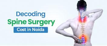
The thoracic spine is the central part of the human spinal column, located between the cervical (neck) and lumbar (lower back) areas. These parts of the spine are responsible for stability, mobility, and guarding organs. For maintenance of the spine and timely medical intervention, it is crucial to know about its anatomy, function, associated conditions, diagnosis, and treatment. Finding the best thoracic spine surgery in Noida is crucial, so contact the nearest hospital with the best reputation in your area and set up your consultation right away.
Book your consultation by calling +91 9667064100 with a spine specialist today and take the first step toward relief.
There are 12 vertebrae in the thoracic spine, labeled T1-T12. It starts at the end of the neck and goes up to just below the lumbar spine, constituting the upper and middle back. They protect the spinal cord and support the rib cage. The thoracic spine is less flexible than the cervical or lumbar spine, thus less prone to injury; however, it can still develop disorders.
Each thoracic vertebra is connected to a rib pair, making up a rigid thoracic cage that protects the heart and lungs. There is an intervertebral disc located between each vertebra, acting as a shock absorber, offering flexibility. Facet joints in this area control and limit spinal movements, enabling proper functioning.
The thoracic spine has some important roles:
Protection: It surrounds the spinal cord and shields nerves that report to organs like the lungs, heart, liver, and intestines.
Support: It stabilizes the rib cage and upper portion to support posture and movement.
Respiration: The configuration of the thoracic area and muscles that are connected to it makes breathing possible by opening up and closing the chest.
Mobility and Stability: While the thoracic spine has little flexion and extension, it also has the greatest rotational motion of any of the spinal segments with stability.
Postural Alignment: It is also responsible for maintaining the normal curve of the spine, which assists with balance and the transmission of forces effectively.
Twelve pairs of spinal nerves exit the thoracic spine:
T1-T2: The nerves impact the upper chest and arms. The T1 nerve is one of the nerves that belong to the brachial plexus, responsible for controlling hand and arm movement.
T3-T5: Regulate chest wall, lungs, and diaphragm.
T6-T12: Contribute support to abdominal and back muscles and help in balance, posture, and actions such as coughing and breathing.
These nerves transmit impulses to and from large organs such as the lungs, heart, liver, kidneys, and small intestine.
While injuries to the thoracic spine are less common given its stiffness, there are still various conditions that will impact it:
Muscle Strain or Irritation: Most often caused by poor posture or prolonged sitting.
Ligament Sprain: Due to sudden twisting motions or trauma.
Spinal Tumors: Benign or malignant tumors that often present as deep, chronic pain at night.
Vertebral Compression Fractures: Most common in elderly or osteoporotic individuals, with resultant pain and loss of height.
Overuse Injuries: Due to repetitive use like lifting, twisting, or bending.
Spinal Fractures: Typically secondary to trauma or bone weakening by osteoporosis or tumors.
With age or wear and tear, the thoracic spine can develop the following:
Spondylosis (Osteoarthritis): Cartilage wear and tear leads to pain and stiffness.
Degenerative Disc Disease: Discs that separate the vertebrae dehydrate and become less elastic.
Spinal Stenosis: Narrowing of the spinal canal that may crush the spinal cord and nerves.
Kyphosis: Excessive forward spinal curve that causes slouching posture.
Herniated Discs: It might occur in the thoracic spine and may press on spinal nerves.
Myelopathy: Compression of the spinal cord that impacts mobility and coordination.
Thoracic nerve damage may lead to:
Radiating pain or numbness closer to the chest or belly
Weakness of limbs or core muscles
Shortness of breath or difficulty breathing
Loss of bowel or bladder control
Decreased sensation in arms, legs, or genitalia
Stiffness or spasms of the muscle
If you do experience these symptoms, ensure you see the best spine surgeon in Noida as soon as possible.
The accurate diagnosis begins with the history and physical examination. The ideal physician can recommend:
X-rays: To observe bone alignment and breaks.
MRI: To examine the spinal cord, discs, and nerve roots.
CT Scan: Gives detailed pictures of vertebrae and structural issues.
Myelogram: Injects contrast dye into the spinal cord and nerves, highlighting them.
EMG and Nerve Conduction Studies: Test for nerve function and identify locations of compression.
Treatment differs by severity and cause. The most common options are:
Physical Therapy: Trims and adjusts posture.
Medications: Muscle relaxants, pain medications, and anti-inflammatories.
Epidural Steroid Injections (ESIs): End nerve pain and decrease inflammation.
Bracing: Helps stabilize the spine when it has fractures or kyphosis.
Decompression Surgery: Removes bone or tissue pressing on nerves.
Spinal Fusion: Merges the spine by rods, screws, and bone grafts.
Tumor Removal: Requires delicate surgical procedures to eliminate tumors from the spine.
The best thoracic spine hospital in Noida has an expert who will prescribe the optimal course depending on the health and the complexity of the condition.
Recovery could include rehabilitation therapies such as
Physical Therapy: Restores strength and mobility.
Occupational Therapy: Helps to adapt to daily routines.
Pain Management: Medication and Non-Invasive Therapies.
Lifestyle Modifications: Posture improvement, weight management, and ergonomic adjustments.
Follow-ups and imaging studies on a regular basis assist in monitoring progress. Follow-up is usually based on the severity of the condition, timely intervention, and patient compliance.
Seek immediate care if you have:
Increasing or persistent back pain
Numbness or weakness in the legs or arms
Loss of bowel or bowel control
Chest pain or shortness of breath
Unexplained weight loss accompanied by back pain
These could be signs of life-threatening conditions like compression, fracture, or tumor in the spinal cord that should be examined immediately at a hospital.
The cost of treating thoracic spine disorders is determined by the following factors:
Diagnostic tests (MRI, CT scan, X-rays)
Treatment process (physical therapy, surgery, medication)
Hospitalization (number of days) and room
Recovery and follow-up after treatment
In India, the cost of treatment ranges from ₹50,000 to ₹5 lakhs or more, depending on how complicated the condition is, the technology used, and the amount of experience of the specialist. Always ask for an estimate from the hospital.
The thoracic spine surgery is instrumental in stabilizing the human body, shielding important organs, and facilitating movement. Pathologies in this area can lead to pain, loss of mobility, or systemic issues if not treated. An early diagnosis, a consultation with a competent spine specialist, and prompt treatment at a well-reputed hospital can significantly enhance the quality of life.
If you’re experiencing persistent upper back discomfort, posture issues, or unusual neurological symptoms, consult a spine specialist today. Prioritize spinal health—get evaluated at the best hospital nearby for expert care and peace of mind.
Q1. Can poor posture alone cause permanent thoracic spine problems?
Ans: Yes, repeated poor posture—especially slouching or forward head position—over time will cause structural changes of the thoracic spine, including kyphosis, muscle imbalance, and premature degenerative disc disease.
Q2. Why does thoracic spine pain worsen at night or with recumbency?
Ans: Thoracic pain increases at night due to decreased distraction of the spinal joints and changes in intervertebral disc pressures. It may also reflect the development of tumors or infections of the spine that require urgent examination.
Q3. Can thoracic spine problems cause heart- or lung-type symptoms?
Ans: Yes. Distressed thoracic nerves can mimic tightness or pain in the chest, commonly mistaken for heart or lung problems. This symptom is very common with thoracic radiculopathy or dysfunction of the costovertebral joint.
Q4. Is exercise with thoracic disc bulging or herniation safe?
Ans: Exercise is generally alright, but twisting or high-impact activity must be avoided without seeking expert guidance. A spine specialist or physiotherapist can come up with a safe regime.
Q5. How do I differentiate thoracic nerve pain from muscular back pain?
Ans: Thoracic nerve pain will radiate around the rib cage in a band-like manner and is sometimes accompanied by generalized tingling or numbness. The pain will generally be localized to the affected area and worsen with specific movements or pressure.
Q6. Can hours of sitting at a desk cause thoracic spine degeneration?
Ans: Yes. Sedentary living and poor ergonomics over time contribute largely to degenerative thoracic changes, especially if compounded by inadequate back and core muscle strength.

Spinal cord injury is one of the most serious injuries one can have. The damages can be permanent if not treated on time.

Advanced neurosurgery improves treatment for brain and spine disorders with better outcomes.

Spinal stenosis surgery is needed when severe pain, weakness, or numbness persists despite non-surgical treatment and affects daily activities.
We all get headaches. Sometimes from stress, dehydration, or staring at a screen too long. But if a headache feels more intense and more sudden, especially followed by confusion, blurry vision, or difficulty speaking, it needs immediate medical attention. It might be a sign of something much more serious like a blood clot in brain.
Therefore, understanding what causes a blood clot, how to spot it, and most importantly, what doctors can do about it, can make all the difference. Timely treatment saves lives – and in many cases, prevents long-term damage.
So if you or a loved one experiences a sudden bout of headaches, confusion, or weakness in your face or limbs, don’t brush it off. These could be early signs of something serious like a brain clot. Seek medical help immediately!
At Felix Hospital, our neurosurgeons are trained to respond quickly with advanced diagnostics and treatment. To schedule an appointment, contact us at +(91) 9667064100!
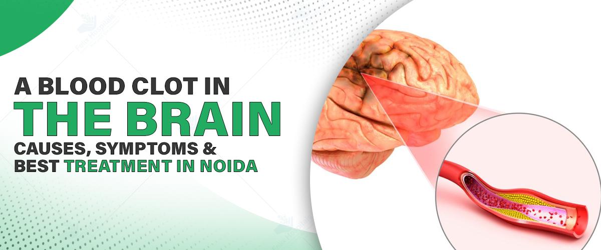
A blood clot in the brain, also known as a cerebral thrombosis or stroke caused by thrombosis, happens when blood flow to part of the brain gets blocked by a clot. The brain, like every other organ, needs oxygen and nutrients. When a clot stops that flow, brain cells can start to die within minutes.
There are different types of strokes caused by clots. The most common is an ischemic stroke, which is due to a blockage. This is different from a hemorrhagic stroke, which involves bleeding in the brain.
Sometimes, the clot forms in a brain artery itself. Other times, it travels from another part of the body, like the heart or neck, and gets stuck in a narrow artery in the brain.
Clots don’t usually form out of nowhere. They often result from other underlying issues. Here are some of the most common causes of brain blood clot:
High blood pressure – Damages blood vessel walls, making clots more likely
Diabetes – Affects how your blood flows and how well your vessels work
Atherosclerosis – Hardening or narrowing of arteries due to plaque buildup
Atrial fibrillation – An irregular heartbeat that can cause clots in the heart to travel to the brain
Smoking – Increases clotting tendency
High cholesterol – Leads to blocked arteries over time
Obesity and sedentary lifestyle – Raise overall risk of stroke and clot formation
Head trauma – In some cases, injury to blood vessels can lead to clot formation
When a blood clot blocks an artery in your brain, symptoms usually come on suddenly. You might feel fine one moment, and very unwell the next. Common signs of blood clot in brain include:
• A sudden, severe headache
• Numbness or weakness, especially on one side of the body
• Difficulty speaking/slurred speech
• Trouble understanding what others are saying
• Blurred or double vision
• Dizziness or loss of balance
• Confusion or trouble concentrating
• Loss of consciousness or seizures
• F – Face drooping
• A – Arm weakness
• S – Speech difficulty
• T – Time to call emergency services
Doctors rely on a combination of symptoms and advanced imaging to figure out what’s going on. The process usually involves:
CT Scan or MRI: These help to identify if there’s a clot or bleeding.
CT Angiography or MR Angiography: These focus on the blood vessels and show exactly where the blockage might be.
Blood Tests: To check clotting factors, sugar levels, and cholesterol
ECG and Echocardiogram: If there’s a chance the clot came from the heart, these tests help detect heart conditions
If you suspect signs of a blood clot in brain, acting fast is the key! The treatment depends on how quickly the person gets to the hospital. The golden window is often the first 3 to 4.5 hours.
Thrombolytic (Clot-Busting) Drugs : The medication works by dissolving the clot and restoring blood flow. It must be given within a few hours of symptom onset to be effective.
Mechanical Thrombectomy: In some cases, especially with larger clots, doctors use a catheter to go into the brain’s arteries and physically remove the clot. This is done under imaging guidance and is highly effective when performed early.
Blood Thinners: If the stroke risk remains high, patients may be put on long-term anticoagulants to prevent future clots.
Lifestyle Changes and Rehabilitation: Post-stroke treatment isn’t just about medication. Physical therapy, occupational therapy, and speech therapy play a big role in recovery, especially if mobility or communication was affected.
A blood clot in the brain is serious and should be treated as a medical emergency. In many cases, it’s preventable. As mentioned, rapid treatment can dramatically change the outcome. At Felix Hospital, we provide comprehensive treatment for brain blood clots, including emergency care. We also provide rehabilitation and support care to all our patients recovering from the after effects of the brain blood clot or brain stroke.
Don’t ignore early signs and symptoms, especially if your body is giving you warning signs. Consult the best neurologists in Noida immediately to seek appropriate treatment.
Q1. Can you survive a brain clot?
Ans: Yes, many people survive and recover well, especially with early treatment. The key is recognizing the signs and getting help fast.
Q2. How long is the recovery after a brain clot?
Ans: It varies. Some recover in weeks, while others may need several months of rehab. Age, overall health, and how quickly treatment was received all play a role.
Q3. Are blood clots in the brain preventable?
Ans: Often, yes. Managing blood pressure, quitting smoking, staying active, and keeping diabetes under control can significantly lower your risk.
Q4. What’s the difference between a stroke and a brain clot?
Ans: A stroke caused by a clot is called an ischemic stroke. So technically, a brain clot is one type of stroke. There are other kinds too, like hemorrhagic strokes, which involve bleeding.

ब्रेन स्ट्रोक एक जानलेवा स्थिति है जिसमें मस्तिष्क को रक्त आपूर्ति रुक जाती है। इसके प्रकार, कारण, लक्षण, बचाव और इलाज

A concise guide explaining brain tumor surgery cost in Noida, including key factors, treatment options, and expert care.

Recognizing the symptoms of a brain stroke is critical for getting prompt medical attention. The acronym F.A.S.T. is often used to remember the most common Brain stroke symptoms.

Brain-boosting foods rich in antioxidants, omega-3s, and vitamins support memory, protect brain cells, and may help reduce the risk of dementia and Alzheimer’s disease.
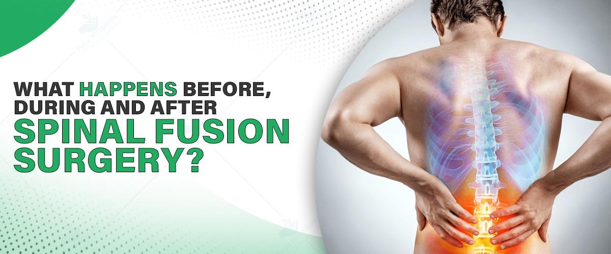
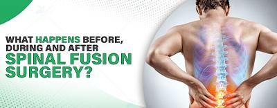
Spinal fusion surgery is a standard process applied to correct different spine disorders like herniated disks, spinal instability, scoliosis, or degenerative disc disease. Through spinal fusion surgery, the goal is to attach two or more vertebrae in your back permanently, removing any motion between them to avoid pain and enhance stability. If you are looking for the best spinal fusion physicians near me, learning about this surgery in detail will help you make informed choices. This blog will prepare you for spinal fusion surgery by explaining what to expect.
Meet the best orthopedic surgeon at the start. Call us now at +91 9667064100.
1. First Consultation and Assessment: The process starts with an initial consultation with a spine expert. The spine expert will assess your symptoms, medical history, and imaging studies such as X-rays, MRIs, or CT scans to decide whether spinal fusion is appropriate for your case. The spinal fusion operation price in Noida varies based on the level of complexity and hospital amenities.
2. Preoperative Instructions: The following suggestions may be included in the preoperative instructions:
Quit smoking, as it affects bone healing.
Avoid taking anti-inflammatory medications that can lead to increased bleeding.
Make arrangements for assistance at home while you are recovering.
Get the required blood work and physical examination.
3. Pre-Surgery Preparation One day before the surgery
You may be requested to:
Fast for a minimum period of 8 hours before surgery
Take a shower using antibacterial soap
Pack a hospital bag with essentials for your hospital stay, such as your medical records and comfortable clothes
1. Anesthesia and Positioning: The surgery is done under general anesthesia. Depending on the part of your spine being operated on, you will lie on your stomach or back.
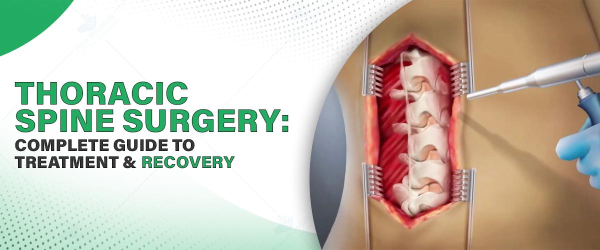
2. The Surgical Process
The surgeon makes incisions over the affected area.
The surgeon removes any ruptured disc material.
A bone graft (either from your body or a donor) is inserted in between vertebrae.
Metal rods, screws, or plates might be attached to stabilize the spine.
Spinal fusion surgery can last several hours, depending on how many vertebrae are fused.
1. Hospital Stay and Recovery Room After surgery: You will be taken to the recovery room, where your vital signs are monitored closely. Most patients are hospitalized for 2 to 4 days. In this process, doctors monitor pain management and the resumption of mobility safely.
2. Mobility and Pain Management
Pain medications are administered to reduce discomfort.
Physical therapists can get you moving within 24 hours.
Gradual walking and basic movements are encouraged to prevent stiffness.
3. Healing and Long-Term Recovery
The fusion process typically takes 6 to 12 months.
You should avoid bending, twisting, or lifting heavy objects.
We may provide a back brace for additional support.
Follow-up visits are necessary to monitor healing and make adjustments in care.
As with any major surgery, spinal fusion comes with certain risks, including:
Infection
Nerve damage
Incomplete fusion (pseudoarthrosis)
Pain at the bone graft site
Talking to your specialist about these risks will allow you to balance the advantages against possible complications.
Decreases chronic back pain
Stabilizes the spine
Enhances mobility and posture
Allows patients to go back to regular activities with improved comfort
Most patients can return to work and physical activities within a few months with proper rehabilitation and medical attention.
Finding the correct team for your treatment is critical. If you are in search of a spinal fusion hospital in Noida, keep the following in mind:
Surgeon experience
Presence of high-end surgical equipment
Facility reputation
Transparency of charges and patient feedback
Selecting treatment at the best Orthopedic hospital in Greater Noida provides you with multidisciplinary teams, post-operative services, and custom treatment plans.
Schedule an appointment with the best doctor in Noida for personalized care and post-operative care.
Spinal fusion is an established way to treat long-term spinal disorders and enhance the quality of life. Be it assessing your options or getting ready for the surgery, having the proper information at hand can assist you in going through the process with ease. If you are looking to undergo the procedure, begin by taking advice from experts at the best hospital in Greater Noida.
From diagnosis to recovery, skilled care ensures you receive the treatment, protection, and support you require for your healing journey.
Q1. What is the recovery time from spinal fusion surgery?
Ans: Recovery can take between 3 to 12 months, depending on patient health, age, and activity level. Physical therapy and routine follow-up are important aspects of recovery.
Q2. Is spinal fusion surgery painful?
Ans: Post-surgery pain is normal, but it is well-managed with medication and improves over time.
Q3. Can spinal fusion fail?
Ans: Infrequently, but sometimes, pseudoarthrosis or non-union occurs, where the two bones do not join together properly. This can be minimized with great surgical skill and good postoperative care.
Q4. Will I become less mobile in my back after spinal fusion?
Ans: Spinal fusion restricts motion between two fused vertebrae but generally does not limit overall mobility. Most patients adjust easily.
Q5. What is the average cost of spinal fusion surgery in Noida?
Ans: It varies with complexity, hospital, and fees of the surgeon. It usually ranges between ₹1.5 lakhs and ₹4 lakhs. Always seek a proper estimate before going for surgery.
Spinal stenosis surgery is needed when severe pain, weakness, or numbness persists despite non-surgical treatment and affects daily activities.
A spinal cord injury occurs when there is damage to the spinal cord, often resulting from trauma, disease, or degenerative conditions.
Spinal fusion surgery is a procedure that joins two or more vertebrae in the spine to reduce pain, correct deformity, or improve stability.
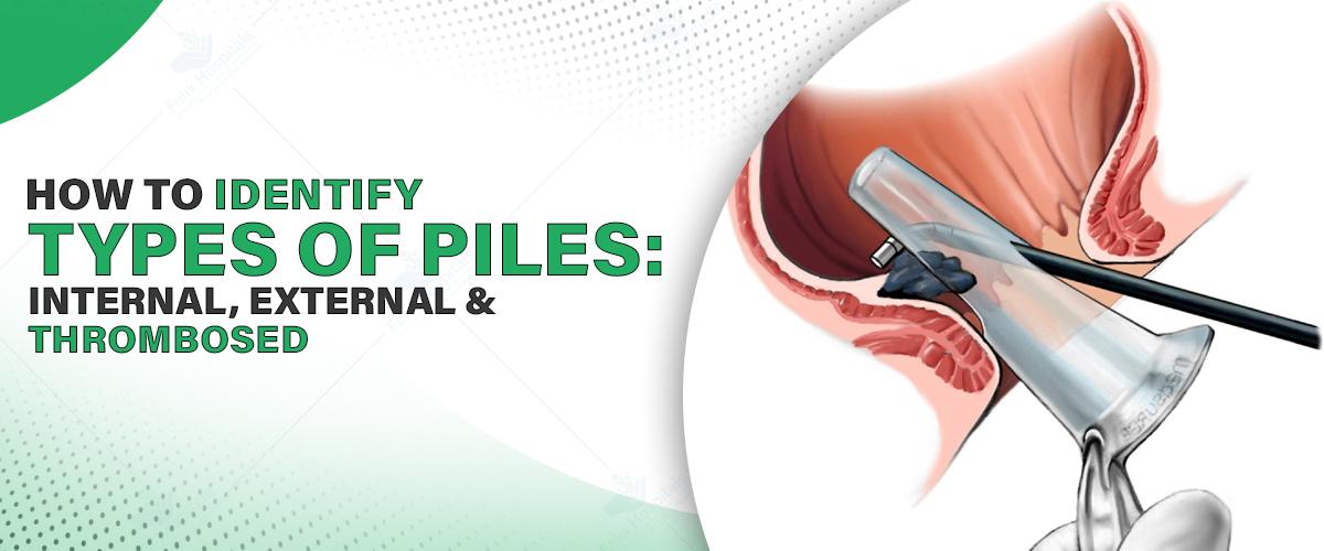
Piles, or hemorrhoids, are a common but rather painful condition that affects the majority of the population, particularly adults older than 30. They develop when the veins in and around the anus or the lower rectum become inflamed or swollen. Though they might not be fatal, they can greatly impair the quality of life unless they are treated at the right time. Familiarity with the various types of piles is the stepping stone for effective management of them.
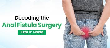
In this detailed blog post, we will help you in determining the three most common types of piles: internal, external, and thrombosed. Each will have different symptoms and treatment procedures. Early identification can aid you in getting proper care at the right time. If you have symptoms, consulting the best hospital for piles treatments can help you get an expert diagnosis of the piles symptoms and treatment specific to your condition.
Consider scheduling a consultation with a specialist today by calling +91 9667064100 to receive expert advice on managing piles effectively.
Piles are inflamed veins found in the lower rectum or anus. They can be developed either internally (within the rectum) or externally (outside the skin near the anus). Piles are caused by several reasons, including prolonged sitting, straining while passing stool, constipation, pregnancy, or obesity. We can categorize them into three types on a broad level:
Internal Piles
External Piles
Thrombosed Piles
Let's study each of them separately.
Internal piles develop within the rectum and are not visible or palpable in the initial phases. As there are no pain-sensing nerves in this region, internal piles do not lead to pain unless they are large or prolapse.
Symptoms:
Bleeding in the rectum without pain either with or after a bowel movement
Discharge of mucus
Feeling of incomplete emptying
Prolapse: In severe cases, internal piles protrude outside the anus and bring about pain or aching resulting in prolapse.
Grade I: No prolapse, just bleeding
Grade II: Prolapse on defecation but spontaneously retraces
Grade III: Prolapse that needs to be manually reduced
Grade IV: Prolapsed piles which cannot be pushed inside
Diagnosis: We generally do an anoscope or proctoscopy to look at internal piles. If the bleeding is chronic, you may have to undergo additional tests such as a colonoscopy to exclude other pathology.
Lifestyle and dietary modifications
Medications and topical therapy
Minimally invasive treatments such as rubber band ligation or infrared coagulation
External piles exist beneath the skin in the area surrounding the anus and are usually visible as lumps. They have more pain-sensitive nerves, so they can be painful, particularly on sitting or passing stools.
Pain or tenderness around the anal area
Itching or irritation
Swelling or lumps that are painful to touch
Minor bleeding while passing stool
Diagnosis: External piles are often diagnosed by a physical examination. The physician can easily check the area to determine the size and severity of the hemorrhoids.
Over-the-counter creams or ointments
Painkillers
Sitz baths (warm water baths for buttocks and hips)
Surgical excision in severe situations
A thrombosed pile is an external pile where there is a blood clot within the hemorrhoidal vein. This creates a hard, tender lump around the anus and can necessitate urgent medical care.
Sharp and severe pain
Inflammation and swelling
Hard, bluish lump close to the opening in the anus
Possible bleeding if the skin is broken
Diagnosis: The thrombosed piles can usually be diagnosed with a visual inspection. Your physician will check the size of the clot and decide on treatment.
Cold compresses and analgesics to alleviate swelling
In certain situations, the physician might do a small procedure to drain the clot.
Surgery in case of recurring symptoms
Several risk factors could result in piles:
Chronic diarrhea or constipation
Straining during defecation
Prolonged sitting, particularly on the toilet
Low-fiber diet
Obesity
Pregnancy (because of enhanced pressure in pelvic veins)
Aging
Family history
Diagnosis typically starts with a physical examination and history taking. Other diagnostic methods are:
Digital rectal exam
Anoscopy or sigmoidoscopy
Colonoscopy (in the event of rectal bleeding or suspected other conditions)
Early diagnosis is important. If you have any of the above symptoms, it's best to seek consultation from the best piles surgery doctors in Noida for proper assessment and treatment.
Lifestyle modifications: Fiber diet, water intake, and avoiding straining
Medications: Analgesics, laxatives, anti-inflammatory ointments
Minimally invasive procedures: Rubber band ligation, sclerotherapy, infrared coagulation
Hemorrhoidectomy: Surgical excision of piles, typically for severe or frequent cases
Stapled hemorrhoidopexy: Staples are applied to excise or reposition piles
Thrombectomy: For thrombosed piles, surgical evacuation of the clot
Although piles are not always preventable, some preventive steps can significantly lower the risk:
Consume a high-fiber diet (whole grains, fruits, vegetables)
Drink adequate fluids
Avoid sitting for long periods
Stay physically active
Empty the bowel as soon as possible
Maintain healthy body weight
Don't neglect signs such as:
Chronic bleeding during defecation
Severe or sharp pain
Hard masses near the anus
Distinct bowel habit changes
Early treatment ensures a better prognosis. Experts at the best pile treatment hospital in Noida provide advanced diagnostic tools and comprehensive care for effective pile management.
Call now at +91 9667064100 and make your first move toward relief. Contact the best hospital in Noida and schedule your consultation today!
Piles are a manageable condition, especially when identified early and treated appropriately. Knowing the differences between internal, external, and thrombosed piles helps in recognizing the symptoms and seeking timely care. Whether you’re dealing with occasional discomfort or a more serious form of hemorrhoids, seeking professional medical advice can significantly improve your comfort and recovery.
Always give importance to your health and consult a specialist if you think you are suffering from piles. A proper diagnosis and treatment by skilled professionals at the top hospital in Noida can provide you with relief, confidence, and overall well-being.
Q1. Will piles resolve on their own without treatment?
Ans: Mild piles can be benefited by some simple lifestyle modifications such as a high-fiber diet and liquids. But more serious piles are usually treated by medicine or surgery.
Q2. Is bleeding the sole sign of piles?
Ans: No. Bleeding is a frequent symptom, but piles may also produce itching, pain, discomfort, swelling, or a lump in and around the anus, depending upon the severity and type.
Q3. Can I treat piles at home without consulting a doctor?
Ans: These may be treated at home with over-the-counter creams, S itz baths, and dietary adjustments. However, in case of continuous, increasing, or heavy bleeding, medical treatment is required.
Q4. Are piles and anal fissures the same?
Ans: No. Piles are inflamed veins, whereas anal fissures are minute tears in the skin around the anus. Though they are caused by and treated differently, piles and anal fissures can have the same symptoms, like pain and bleeding.
Q5. What is the quickest method to alleviate pain from thrombosed piles?
Ans: Using cold compresses, anti-inflammatory creams, and pain relievers are some ways it can give short-term relief. In other instances, a little surgery might be needed to have the blood clot removed for instant relief.

Know the symptoms of piles in females, including discomfort, bleeding, and swelling, along with causes and treatment options for relief.

Dr. Ritesh Kumar Agrawal is the Best Doctor for piles treatment in Noida at Felix Hospital. With years of specialized experience in proctology.

वल्वर कैंसर महिलाओं में होने वाला एक दुर्लभ लेकिन गंभीर कैंसर है, जो महिलाओं के जननांग के बाहरी हिस्से (वल्वा) को प्रभावित करता है। समय पर पहचान और इलाज से हम जीवन की गुणवत्ता को बनाए रख सकते है। अगर आपको भी अपने या अपने प्रियजन के अंदर कुछ बदलाव या लक्षण दिख रहे हैं तो समय रहते ग्रेटर नोएडा में सर्वश्रेष्ठ स्त्री रोग अस्पताल (Best Gynecology Hospitals in Greater Noida) से सलाह प्राप्त कर सकते है। इस ब्लॉग पोस्ट में हम जानेंगे कि वल्वर कैंसर क्या होता है?, इसके लक्षण, कारण, जांच और इलाज गाइनोकोलॉजिकल गाइडलाइन्स के अनुसार कैसे किए जाते हैं।
ज्यादा जानकारी के लिए हमें कॉल करें +91 9667064100.
वल्वर कैंसर (Vulvar cancer) महिलाओं के बाहरी जननांगों यानी वल्वा में विकसित एक दुर्लभ कैंसर है। यह कैंसर वल्वा की त्वचा या इसके नीचे की कोशिकाओं में असामान्य रूप से बढ़ता है। जिससे ट्यूमर होता है। यह कैंसर धीरे-धीरे होता है। अगर समय रहते पहचान न हो तो यह आसपास के अंगों में भी फैल जाता है।
वल्वा महिलाओं की प्रजनन प्रणाली का बाहरी हिस्सा है। जिसमें लैबिया माजोरा (बाहरी होंठ), लैबिया मिनोरा (भीतरी होंठ), क्लिटोरिस, वेस्तिब्यूल (योनि का प्रवेश द्वार), यूरिन मार्ग का द्वार (यूरेथ्रा ओपनिंग) ये मुख्य अंग शामिल होते हैं। वल्वा महिला के जननांगों की पहली सुरक्षा परत होती है। यह यौन स्वास्थ्य व यूरिन संबंधी कार्यों में मुख्य भूमिका निभाती है। यह कैंसर अक्सर वृद्ध महिलाओं में दिखता है। मगर कुछ मामलों में यह कम उम्र की महिलाओं को भी प्रभावित करता है। खासकर अगर एचपीवी (एचपीवी) संक्रमण मौजूद हो।
यह सबसे सामान्य प्रकार है लगभग 70%–90% मामलों में होता है। यह वल्वा की बाहरी त्वचा की परत की कोशिकाओं में होता है।
यह त्वचा के रंग बनाने वाली कोशिकाओं (मेलानोसाइट्स) से शुरू होता है। यह तेजी से फैलता है। अक्सर गहरे रंग के धब्बे या गांठ के रूप में प्रकट होता है।
यह बहुत धीरे बढ़ने वाला कैंसर है। वल्वा पर यह बहुत कम दिखता है। लेकिन स्किन कैंसर (skin cancer) के रूप में मौजूद होता है।
यह ग्रंथि कोशिकाओं से उत्पन्न होता है और दुर्लभ प्रकार में गिना जाता है। यह तब होता है जब ग्रंथियों की कोशिकाएं असामान्य रूप से बढ़ती हैं।
यह वल्वा के गहरे टिशू से उत्पन्न होता है और अत्यंत दुर्लभ होता है। यह सामान्यतः सॉफ्ट टिशू सारकोमा की श्रेणी में आता है।
वल्वर कैंसर का कोई एक निश्चित कारण नहीं होता, लेकिन कुछ मुख्य जोखिम कारक और संभावित कारण निम्न हैं:
ह्यूमन पैपिलोमावायरस (एचपीवी) एक आम यौन संचारित वायरस है। जो वल्वर कैंसर में पाया जाता है। विशेषकर एचपीवी-16 और एचपीवी-18 प्रकार कैंसरकारी होते हैं। जो लंबे समय तक एचपीवी संक्रमण रहने पर वल्वा की कोशिकाओं में कैंसरजनक बदलाव होता हैं।
वल्वर कैंसर वृद्ध महिलाओं में अधिक देखा जाता है। 60 वर्ष से अधिक आयु की महिलाओं में इसका जोखिम अधिक होता है। उम्र के साथ कोशिकाओं की मरम्मत की क्षमता कम होती है।
जिन महिलाओं की इम्यून सिस्टम कमजोर होता है। यानी एचआईवी/एड्स (HIV/AIDS) या ऑर्गन ट्रांसप्लांट लेने वाली महिलाओं में कैंसर से लड़ने की क्षमता घटती है। एचपीवी जैसे वायरस भी ऐसे शरीर में सक्रिय रहते हैं।
धूम्रपान करने वाली महिलाओं में एचपीवी संक्रमण के कैंसर में बदलने का खतरा अधिक होता है। निकोटिन व अन्य टॉक्सिन्स से शरीर की कोशिकाओं को नुकसान होता है।
प्री-कैंसर की इस स्थिति में वल्वा की कोशिकाएं असामान्य होती है। समय रहते वीआईएन का इलाज न होने यह वल्वर कैंसर में बदलता है। यह स्थिति विशेष रूप से एचपीवी पॉजिटिव महिलाओं में दिखती है।
यह एक दीर्घकालिक त्वचा रोग है, जो वल्वा की त्वचा को पतला, सफेद और खुजलीदार बनाता है। समय के साथ यह त्वचा कैंसर (skin cancer) की संभावना को बढ़ाता है। खासकर बुजुर्ग महिलाओं में होता है।
शुरुआती लक्षणों को अगर समय पर पहचान जाए तो इलाज संभव है। अक्सर महिलाएं इन लक्षणों को नजरअंदाज करती हैं, जो बाद में गंभीर रूप लेती है।
खुजली या जलनः
वल्वा में बार-बार या लंबे समय तक होने वाली खुजली एक प्रमुख संकेत है। यह साधारण इन्फेक्शन होता है। जब यह लगातार हो तो सावधानी जरूरी है।
दर्दः
पेशाब करते समय व चलने के दौरान दर्द महसूस होता है। यह दर्द कभी हल्का और कभी तेज होता है।
रक्तस्रावः
रजोनिवृत्ति के बाद प्राइवेट पार्ट से खून आना चिंताजनक संकेत है साथ ही इसमें बिना कारण के डिस्चार्ज या दाग-धब्बे भी दिखते हैं।
वल्वा में गांठ:
वल्वा की सतह पर कोई कठोर गांठ, मस्सा या न भरने वाला छाला दिखाई दे तो तुरंत जांच करानी चाहिए, ये घाव दर्दरहित भी होते हैं।
त्वचा में रंग का परिवर्तनः
वल्वा की त्वचा का रंग सफेद, गहरा या धब्बेदार हो जाता है। त्वचा मोटी, खुरदरी या पपड़ी जैसी होना भी कैंसर का संकेत है।
शुरुआती लक्षण बहुत हल्के होते हैं। इन्हें नजरअंदाज करना खतरा बढ़ाता है। 60 वर्ष से अधिक उम्र की महिलाओं और एचपीवी संक्रमित महिलाओं को सतर्कता बरतनी चाहिए, जिसके लिए उन्हें समय पर स्त्री रोग विशेषज्ञ से सलाह लेना आवश्यक है।
वल्वर कैंसर की पहचान और इलाज के लिए शुरुआती जांच जरूरी है।
शारीरिक जांचः
डॉक्टर मरीज के वल्वा क्षेत्र की जांच करते हैं और त्वचा की बनावट, रंग, गांठ, सूजन, घाव देखते हैं साथ ही लिंफ नोड्स में सूजन की जांच करते हैं, जिससे कैंसर के फैलाव का संकेत पता चलता है।
बायोप्सी:
बायोप्सी वल्वर कैंसर की जांच के लिए महत्वपूर्ण टेस्ट है। इसमें वल्वा के संदिग्ध भाग से छोटा सैंपल निकालकर प्रयोगशाला में जांच को भेजा जाता है, जिससे कैंसर के प्रकार और ग्रेड को समझने में मदद मिलती है।
कोलपोस्कोपीः
इस माइक्रोस्कोपिक जांच में वल्वा की कोशिकाओं को जूम कर देखते हैं। कोलपोस्कोपी बायोप्सी से पहले होने वाली सहायक जांच है।
इमेजिंग टेस्टः
अगर बायोप्सी में कैंसर की पुष्टि हो जाती तो यह जानना जरूरी होता है कि कैंसर कहां तक फैला है इसके लिए यह टेस्ट जरूरी होते हैं। एमआरआई में वल्वा और आसपास के ऊतकों की स्पष्ट तस्वीर मिलती है। सीटी स्कैन में शरीर में कैंसर के फैलाव, विशेषकर लिंफ नोड्स और पेट के अंगों में जानकारी देता है। पेट स्कैन में सक्रिय कैंसर कोशिकाओं का पता चलता है।
वल्वर कैंसर को उसके फैलाव और गंभीरता के आधार पर चार चरणों में वर्गीकृत किया जाता है।
स्टेज 1 में कैंसर केवल वल्वा तक सीमित रहता है। ट्यूमर का आकार दो सेंटीमीटर से छोटा होता है।
स्टेज 2 में कैंसर वल्वा से बाहर की त्वचा या ऊतकों में फैलता है। यह लिम्फ नोड्स तक नहीं पहुंचता।
स्टेज 3 में कैंसर जांघ के पास स्थित लिम्फ नोड्स में फैल जाता है।
स्टेज 4 में कैंसर यूरेटरी, मलाशय या शरीर के अन्य दूरस्थ अंगों जैसे फेफड़ों तक फैलता है।
इलाज का चयन ट्यूमर के आकार, स्थान, स्टेज और मरीज की शारीरिक स्थिति के आधार पर किया जाता है।
सर्जरीः
वल्वर कैंसर के इलाज के तरीकों में सर्जरी पहली और सबसे महत्वपूर्ण लाइन ऑफ ट्रीटमेंट होती है, इसमें ट्यूमर और उसके आसपास की स्वस्थ ऊतक को सुरक्षित अंतर के साथ हटाया जाता है। वल्वेक्टोमी में कैंसरग्रस्त भाग हटाया जाता है वहीं पूर्ण वल्वेक्टोमी में जब कैंसर फैला हो, तब पूरी वल्वा हटानी पड़ती है। लिम्फैडेनेक्टॉमी में लिम्फ नोड्स में कैंसर फैलने की संभावना हो तो यह जांच और उपचार आवश्यक होता है।
रेडियोथेरेपीः
रेडियोथेरेपी में उच्च ऊर्जा वाली किरणों से कैंसर कोशिकाओं को नष्ट किया जाता है। जब ट्यूमर पूरी तरह हटाना संभव न हो या ऑपरेशन के बाद बचे हुए कोशिकाओं को खत्म करने के लिए होता है। अगर लिम्फ नोड्स में कैंसर फैल गया हो तो रेडियोथेरेपी जरूरी होती है। रेडियोथेरेपी सर्जरी से पहले या बाद में दी जा सकती है।
कीमोथेरेपीः
कीमोथेरेपी में कैंसर कोशिकाओं को मारने के लिए दवाओं का इस्तेमाल किया जाता है। खासतौर से जब कैंसर एडवांस स्टेज में हो या पुनरावृत्ति की संभावना हो। इसमें कई बार रेडियोथेरेपी (Radiotherapy) के साथ कीमोथेरेपी को एक साथ दिया जाता है। इससे रेडिएशन का प्रभाव और अधिक बढ़ता है। कीमोथेरेपी के दुष्प्रभावों में बाल झड़ना, कमजोरी, मतली हो सकते हैं।
इम्यूनोथेरेपीः
नई रिसर्च आधारित इलाज जो उन्नत या मेटास्टेटिक वल्वर कैंसर के लिए उपयोगी हो सकते हैं। यह इलाज अभी क्लिनिकल ट्रायल या सीमित उपयोग में हैं, खासकर जब पारंपरिक इलाज बेअसर हो।
वल्वर कैंसर के इलाज की योजना मुख्य रूप से इस पर निर्भर करती है कि कैंसर किस चरण में है।
स्टेज 1: लो-रिस्क ट्यूमर और ट्रीटमेंटः
ट्यूमर छोटा होता है और वल्वा तक ही सीमित रहता है, लिम्फ नोड्स में फैलाव नहीं होता। यदि ट्यूमर की गहराई कम हो और किनारों में कैंसर न हो तो कोई अन्य उपचार आवश्यक नहीं। सेंटिनल लिम्फ नोड बायोप्सी किया जा सकता है यदि ट्यूमर 1 mm से गहरा हो।
स्टेज 2: सर्जरी के साथ या बिना रेडियोथेरेपीः
ट्यूमर वल्वा से आगे आसपास के ऊतकों (जैसे यूरीथ्रा, वेजाइना, एनस) में फैल चुका होता है, लेकिन लिम्फ नोड्स में नहीं गया होता। यदि ट्यूमर का साइज बड़ा है या मार्जिन क्लीन न हों तो रेडियोथेरेपी दी जाती है, इसमें लिम्फ नोड एसेसमेंट जरूरी है।
स्टेज 3-4: मल्टीमॉडल थेरेपीः
इसमें कैंसर लिम्फ नोड्स तक फैल चुका होता है। यह यूरीनरी ब्लैडर, मलाशय, या शरीर के दूरस्थ अंगों तक फैलता है।
अगर कैंसर वापस लौट आए या स्टेज 4 बी में स्थानीकृत हो और अन्य इलाज विफल हो जाए। पेल्विक एक्सेंटेरेशन एक बड़ी सर्जरी होती है। जिसमें वल्वा के साथ साथ मूत्राशय, यूटेरस, वेजाइना और/या मलाशय को भी हटाया जा सकता है। यह प्रक्रिया तब अपनाई जाती है जब रोगी शारीरिक रूप से सक्षम हो और कैंसर सिर्फ पेल्विक क्षेत्र तक सीमित हो।
वल्वर कैंसर का इलाज समाप्त होने के बाद भी ध्यान देना जरूरी होता है, क्योंकि यह रोग शरीर के साथ-साथ मानसिक और भावनात्मक स्तर पर भी गहरा असर डाल सकता है।
वल्वा की सर्जरी या रेडियोथेरेपी के बाद त्वचा में कठोरता, सूखापन या जलन हो सकती है। महिला की सेल्फ इमेज, आत्मविश्वास और यौन पहचान प्रभावित हो सकती है। निराशा, चिंता, डिप्रेशन जैसी समस्याएं आ सकती हैं। गाइनोकोलॉजिकल ऑन्कोलॉजी सोसाइटीज द्वारा निर्धारित समयबद्ध फॉलोअप बहुत जरूरी होता है। वल्वर कैंसर में पहले 2–3 वर्षों में रिकारेंस की संभावना अधिक रहती है।
गाइनेकोलॉजिकल ऑन्कोलॉजिस्ट मुख्य विशेषज्ञ जो स्त्री जननांग अंगों (जैसे वल्वा, गर्भाशय, अंडाशय) के कैंसर के निदान और इलाज में प्रशिक्षित होते हैं। यह सर्जरी, कीमोथेरेपी और इलाज की योजना यही डॉक्टर बनाते हैं। अगर गाइनेकोलॉजिकल ऑन्कोलॉजिस्ट उपलब्ध नहीं हो, तो यह विशेषज्ञ कैंसरग्रस्त ऊतक को शल्य क्रिया द्वारा निकालते हैं।
वल्वर कैंसर समय रहते निदान हो तो इलाज आसाना होता है। क्योंकि इससे शरीर का संवेदनशील हिस्से प्रभावित होता है। इसलिए शारीरिक, भावनात्मक समर्थन जरूरी होता है। गाइडलाइन आधारित उपचार से जीवन की गुणवत्ता में सुधार होता है। एचपीवी वैक्सीनेशन, धूम्रपान छोड़ना और वीआईएन जैसी प्री-कैंसर अवस्थाओं की निगरानी से इसे रोका जा सकता है। महिला को वल्वा संकोच नहीं, सजगता रखनी चाहिए। समय पर जांच, सही उपचार और जागरूकता वल्वर कैंसर से लड़ने का सबसे कारगर रास्ता होता है।

Cervical cancer is most common in women after breast cancer. Cervical cancer starts from the cervix, i.e., the cervix. The cervix is the lower part of the uterus that connects the vagina to the uterus.

Recent studies have explored a possible link between frequent mouthwash use and cancer risk.

Explore essential information about breast cancer, including its symptoms, causes, types, and treatments, to stay informed and proactive about your health.
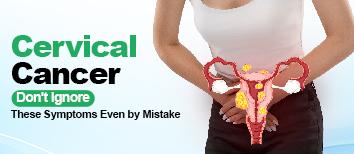
Recognize the early warning signs of cervical cancer. Learn about its symptoms, causes, and when to consult a gynecologist for timely diagnosis and treatment.

Ear infection or otitis media is one of the leading causes for individuals—children primarily—seeing physicians. The condition can result in pain, discomfort, and complications if not treated. It is important to know what causes ear infections, how to recognize the symptoms, and how to treat them to have healthy ears and prevent long-term complications.
In this post, we will discuss why (or "triggers") ear infections occur, what are the various types, who are the most prone to it, and the medications to manage or avoid them.
Protect Your Child's Hearing—Schedule an ENT Check-up in Noida. Call Now at +91 9667064100 or Book an Appointment Online at Felix Hospitals.
Otitis media is inflammation or infection of the middle ear—the air-filled space located behind the eardrum. Otitis media can be due to viruses or bacteria and is likely to arise as a secondary infection from other infections like cold, flu, or respiratory infection that move towards the middle ear.
Three prominent forms of otitis media exist:
Acute Otitis Media (AOM) – an acute infection that causes inflammation, accumulation of fluid, and pain.
Otitis Media with Effusion (OME) – fluid is trapped in the ear after the infection has cleared but no active evidence of infection.
Chronic Otitis Media – persistent or recurring ear infection, which can result in damage to the middle ear and eardrum.
Knowing what triggers ear infections enables it to be prevented or treated early. Here are the main culprits:
Cold, flu, and sinus infections can lead to obstruction and swelling of the Eustachian tubes—the tubes that link the middle ear to the back of the throat. When they are obstructed, fluid will build up in the middle ear, providing a setting where viruses or bacteria can multiply.
Seasonal or environmental allergies may also produce inflammation of the nasal passages and Eustachian tubes. Inflammation raises the risk of fluid accumulation and infection.
Children are especially susceptible to ear infection because their Eustachian tubes are shorter and more horizontal. This allows infections to travel more easily to the middle ear. Their immune systems also are not yet mature, so they are more likely to be infected in general.
Babies who sleep with bottles in their mouths or who use pacifiers all the time might have a higher risk of developing ear infections. It's easier for the fluid and the bacteria to travel into the ear canal when they are in this position.
Secondhand smoke and indoor air pollution will cause irritation to the respiratory tract and Eustachian tubes, which will raise the risk of infection. They are much more likely to have repeated infections in the ear if children smoke.
People with weakened immunity from chronic illness, drugs, or immune dysfunction are at higher risk of repeated bouts of ear infection.
Ear infections occur more frequently during the fall and winter months, mostly because of more colds and flu. Dry indoor air and temperature changes can also be contributing factors.
Early detection of symptoms will enable prompt diagnosis and treatment. The most frequent signs of ear infections are:
Ear pain (mild to severe)
Pulling or tugging on the ear (in children)
Difficulty hearing or reacting to sounds
Drainage of fluid from the ear
Irritability or fussiness in children
Difficulty sleeping
Loss of balance
Ear pressure or headache
In untreated or chronic infections, including ruptured eardrum or hearing loss, complications may arise. If you are facing some issue related to otitis media, simply go to the best ENT hospital near me for the best treatment.
Physicians generally diagnose ear infections using an otoscope, a device that enables them to peer into the ear canal and examine the eardrum for redness, swelling, or fluid. Other tests such as tympanometry or audiometry can be performed in some instances, particularly if fluid accumulation or hearing loss is suspected.
Treatment for Noida ear infection varies according to their type, severity, and patient age.
Doctors can order watchful waiting treatment for children older than two years for minor infections. Most ear infections will get better without the use of antibiotics.
When there are persistent or aggravating symptoms, ampicillin-type antibiotics may be used. One needs to finish the entire course in order not to have recurrence or antibiotic resistance.
Acetaminophen or ibuprofen, over-the-counter medications, can be prescribed to mitigate pain and fever. Always consult a physician at the best ENT hospital in Noida before administering medication to children.
Ear drops could be prescribed in certain situations to ease pain or cure outer ear infections that are part of otitis media.
It is a simple procedure in which an extremely small tube is inserted through the eardrum to drain fluid and reduce pressure. It is used when repeated infections occur or hearing is significantly impacted.
Recurring infections can be prevented by treating underlying conditions such as allergies, clearing allergens from the home, or sinus problems.
It cannot be prevented for all ear infection cases, but there are a number of habits which lower the risk:
Breastfeed children to enhance immunity and prevent the risk of infection.
Don't smoke in the house or around children and expose them to secondhand smoke.
Maintain hygiene like hand washing to avoid cold and flu.
Follow up with vaccination, particularly flu shot and pneumococcal vaccination.
Feed babies in an upright position and not while lying down.
Restrict use of a pacifier, particularly after 6 months.
Treating allergy and congestion in advance.
In case you have symptoms of otitis media, either in kids or yourself, getting the proper diagnosis and on-time treatment from a professional ENT doctor is of extreme importance.
The best ENT experts at Felix Hospital, Noida, offer:
Dr. Arvinder Pal Singh – Experienced in diagnosing and treating complex ear issues through patient-focused approaches.
Dr. Arjun Saini – An extremely experienced doctor in treating chronic and acute ear infections with the most recent ENT procedure and with care.
Felix Hospital Noida has the latest diagnostic equipment and offers complete ENT treatment for all age groups at a reasonable cost in Noida. Don't delay an ear infection—early treatment can avoid complications and lead to healthier ears.
Persistent earache, hearing loss, or frequent infection may be riskier than you imagine. Schedule a consultation with the finest ENT doctors at Felix Hospital, Noida.
Ear infections are a prevalent yet controllable illness, provided that they are diagnosed early and addressed adequately. Having knowledge of the causes of otitis media—either allergy, infection, or anatomy—empowers parents and adults to act beforehand. Should you or your child have signs of ear infections, do not delay in seeing an experienced, seasoned professional ENT specialist with comprehensive evaluation and tailored plan of treatment. Early action not only brings relief from pain but safeguards long-term ear health.
Q1. Can ear infections impact my child's speech development?
Ans: Yes, recurrent or untreated ear infections may cause temporary or permanent hearing impairment, which could result in slowed speech and language development in infants and toddlers.
Q2. How do I determine if the ear infection is bacterial or viral?
Ans: It is difficult to say without a proper check-up. Viral infections, though, generally get better within 48–72 hours, but bacterial infections get worse or stick around and generally need antibiotics. Only an ENT expert can make a diagnosis.
Q3. Can one fly with an ear infection?
Ans: Flying with an active ear infection can aggravate symptoms from pressure changes. It is best to consult an ENT specialist prior to flying if you have ear pain or congestion.
Q4. Why does my child develop ear infections following every cold?
Ans: Having repeated colds can lead to Eustachian tube dysfunction, particularly in children. The tube maintains middle ear pressure, and when it is blocked, it allows for an environment where infections will grow.
Q5. Do allergies increase the risk of ear infection?
Ans: Yes. Allergies can result in nasal congestion and fluid buildup, which can lead to Eustachian tube blockage and increase the risk of development of otitis media.
Q6. What are the outcomes of not treating chronic otitis media?
Ans: Untreated chronic infections can lead to hearing loss, perforated eardrums, or spread of infection to surrounding structures, like mastoid bones or the brain in extreme cases.
Q7. Do grown-ups get otitis media, too, or is otitis media a kids' disease?
Ans: Although more prevalent in kids, adults do get otitis media—particularly if they're smokers, they have allergies, sinus problems, or a poor immune system.

There has been a steep rise in hearing issues in the last 5 years. People who use earphones and headphones continuously for long duration are prone to become deaf.

Search the best hospital for your child in Noida the team of experienced pediatricians is dedicated to supporting parents and guiding children on their journey to a healthy life.

Felix Hospital in Noida is well known for its general practitioners who offer holistic treatment for different health issues, like the common cold and coughs.

Cold sores, also known as fever blisters, are small, fluid-filled blisters that typically appear on or around the lips.
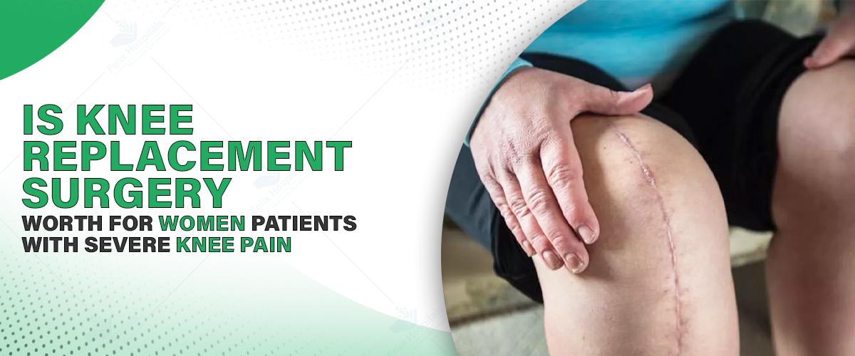
Millions of individuals across the globe have knee pain, which is a prevalent health issue. Injury, arthritis, excess weight, and degenerative joint conditions are just a few of the factors that may lead to it. Both genders can experience knee pain, but research and clinical experience indicate that women tend to experience knee problems more than men. This disparity is due to factors such as variations in hormones, body structure, and way of life.
For many women, knee pain begins as a trivial annoyance but can become an outright problem that makes it difficult to walk, climb stairs, or even remain stand for long periods. When symptoms are not relieved by conservative therapies like medication, physical therapy, and lifestyle changes, knee replacement surgery often becomes a viable and effective option.
This blog post discusses knee replacement surgery in a very detailed manner, focusing on the impact it leaves on women. It also provides advice on how to make an informed decision, specifically for those opting for knee replacement surgery in Noida.
Begin your path to a pain-free life. Schedule a personalized appointment today by calling +91 9667064100.
Replacement surgery for the knee, or knee arthroplasty, is a surgical procedure where the worn-out or broken portions of the knee joint are removed and replaced with man-made parts that are made of metal alloys, ceramics, or high-strength plastic. The reason for this surgery is to relieve pain, restore function, and improve the quality of life for individuals with severe joint disease or damage.
There are various types of knee replacement surgery:
Total Knee Replacement (TKR): Replaces the entire knee joint.
Partial Knee Replacement (PKR): Only the portion of the knee that is fractured is replaced.
Minimally Invasive Surgery: Smaller incisions and quicker recovery.
Advancements in medical technology have quickened recovery, shortened hospital stays, and enhanced surgical success—making knee replacement surgery cost in Noida more economical and accessible. Knee replacement surgery has revolutionized greatly over the years.
Knee issues are more common and severe in women than in men. This added susceptibility can be traced to several main causes:
Women tend to have a broader pelvis than men, which alters the alignment of the femur and tibia. The Q-angle, also known as the quadriceps angle, exerts more pressure on the knee joint, especially during physical activity. Misalignment issues will wear the cartilage down quicker, potentially resulting in conditions such as patellofemoral pain syndrome or osteoarthritis.
Hormonal changes, particularly estrogen, can weaken and stiffen ligaments. Women's joints can be looser at times, such as during their period, pregnancy, or menopause. This predisposes them to injuring their ligaments, such as rupturing their ACL, and accelerates the progression of knee worsening.
Some activities or jobs that women often do might make them more prone to repetitive stress or overuse injuries. In addition, women are more likely to seek a doctor than men, resulting in earlier diagnosis and treatment. The study also indicates how prevalent knee issues are.
Studies prove that women are more susceptible to developing osteoarthritis after 50 years of age. Hormonal changes after menopause can also accelerate joint destruction, which would make surgery a feasible means for long-term relief.
A woman suffering from constant and severe knee pain can transform her life with knee replacement surgery. The following are some of the most significant advantages:
Chronic knee pain can make it difficult to do simple things. Women can resume a more active lifestyle after knee replacement surgery, which can significantly reduce or eliminate pain from arthritis or trauma.
Most say they can do things they couldn't do due to pain, like walking, exercising, or enjoying activities. Improved joint function increases the independence of people.
Being pain-free from chronic pain and more mobile can contribute to your mental well-being, make you sociable, and make you feel healthier in general. Women tend to sleep better, require fewer medications, and are happier about their everyday lives.
Contemporary knee prosthetics last 15 to 20 years, sometimes even longer, under excellent care. This extended lifespan is due to advances in implant material and surgical techniques. As it lasts for a very long period, surgery is an effective long-term remedy.
Most manufacturers currently have gender-specific implants that are designed to fit and feel more like natural tissue, resulting in a better success rate and feel for females. They realize that male and female knees are not the same.
Knee replacement surgery has numerous advantages, but women must think seriously about the risks and duties involved with it. Key factors to consider are:
Recovery from knee replacement surgery takes time, effort, and commitment to physical therapy. It usually takes most patients 6 to 12 weeks to resume their daily lives, but it might take 6 months or more to recover completely.
There are certain complications associated with any major surgery, including
Infection
Blood clots
Nerve damage
How the body responds to anesthesia
Failure or loosening of an implant
Younger women will need to consider how long implants will last and if they will ever have to undergo surgery again in the future. Active individuals will have to learn new patterns of movement and avoid activities that put pressure on the new joint.
If you have to undergo surgery, you need to be strong in mind and have a budget plan. While deciding, you should consider aspects such as insurance coverage, hospital expenditure, and follow-up procedures.
The standard of medical treatment plays a significant role in the effectiveness of knee replacement surgery. If you or a loved one is planning to undergo knee replacement surgery, it is essential to choose the best knee replacement hospital in Noida for this procedure.
One should look for the following points before opting for surgery:
Experienced orthopedic surgeons: Identify hospitals with board-certified orthopedic surgeons with a high volume of knee replacement surgeries. Surgeons with a high volume of successful procedures are typically more skilled at tackling complex cases.
Surgical Facilities of the Future: Newer operating suites equipped with robotic-assisted surgery equipment, 3D imaging, and advanced navigation systems can make the procedure more precise and enhance patient outcomes.
The top hospitals provide comprehensive care services: Diagnosis, individualized treatment planning, physical therapy, dietary counseling, and pain control pre- and post-surgery.
Reputation and Accreditation: Ensure the hospital is accredited by prominent healthcare organizations and has favorable patient reviews and high success rates for joint replacement procedures.
Schedule an appointment with one of the top orthopedic surgeons in Noida to receive a comprehensive examination and a treatment plan made especially for you.
Knee replacement surgery can be a highly effective solution for women suffering from severe knee pain when non-surgical treatments fail. With the use of cutting-edge technologies such as robotic-assisted surgery, computer navigation, and advanced implant materials, the procedure has become safer, more precise, and recovery-friendly. Modern techniques focus on personalized treatment, improved implant alignment, reduced pain, and faster rehabilitation, helping women regain mobility and improve quality of life.
Key Points

Dr. Keshav Goel is an experienced Orthopedic and Joint Replacement Surgeon in Noida with over 10 years of clinical expertise. He has performed 2,000+ independent surgeries and specializes in computer-assisted joint replacement and arthroscopic surgeries of the knee, shoulder, and hip. Known for his skill in treating sports injuries, trauma, and joint disorders, he provides advanced and patient-focused orthopedic care.
For women with long-term and severe knee pain, knee replacement surgery can turn their lives around. It alleviates pain, restores joints to function, and allows you to resume being active. But don't do it impulsively. To make your best decision, you must be aware of the advantages and disadvantages, discuss your individual health requirements with a seasoned orthopedic surgeon, and balance the risks.
If you are considering undertaking this surgery in Noida, it is important that you have the best hospital with experienced surgeons and modernized facilities. A trustworthy medical support system can ease the journey and make the outcome much superior.
If you or someone you love is struggling with knee pain and considering surgery, now is the time to consult a professional. A pain-free future and full mobility are highly probable with the proper assistance and attention.
Q1. Does knee replacement surgery perform equally well for women as it does for men?
Ans: Yes, but typically women receive more pain relief and improved mobility post-surgery. Women-specific implants and surgical methods have enhanced women's success and satisfaction rates.
Q2. What impact do post-menopausal changes in hormones have on the outcomes of knee replacement surgery?
Ans: Women who have undergone menopause can have less dense bones and heal in different ways. Surgeons in the top-ranked hospitals consider these changes when selecting implants and managing patients photoporation, resulting in improved outcomes.
Q3. If I have other illnesses, such as diabetes or thyroid issues, can I still undergo knee replacement surgery?
Ans: Yes, but it's important to monitor other health issues. Both prior to and following the surgery, hospitals in Noida provide multidisciplinary care, including endocrinologists to ensure you have optimal health.
Q4. Is robotic-assisted knee replacement safer or more accurate for women?
Ans: Robotic surgery is more precise, particularly for women with uncommonly shaped joints. It assists in proper alignment, reducing the number of issues and extending the lifespan of implants.
Q5. For how many days do women in Noida usually remain hospitalized after undergoing knee replacement surgery?
Ans: The majority of women are hospitalized for three to five days, which depends on their health and the speed of recovery. All this can be reduced with improved recovery plans and less aggressive methods.
Q6. Will I be able to kneel, squat, or do yoga after knee replacement surgery?
Ans: High-flex implants can allow some individuals to squat and kneel, but this is not the case with all individuals. In three to six months, most women resume low-impact activities such as yoga, swimming, and cycling.
Q7. How do I make the choice between full knee replacement and partial knee replacement?
Ans: This varies depending on how severe the damage is to the joint. Partial replacements are not as invasive and might be a better option for younger women or patients with knee damage restricted to one area. Total replacements are ideal for individuals with more extensive arthritis. Depending on imaging and lifestyle considerations, your orthopedic surgeon will assist you in making the decision.

Gallbladder issues can be a source of major disruption to the day, like painful eating, nausea, or ongoing discomfort. If gallstones or something else are the underlying cause, a cholecystectomy can be the answer. This routine surgery takes out the gallbladder, a small sac under the liver that holds bile to assist with fat digestion. At the top hospital for gallbladder surgery, we aim to guide you through the process of a cholecystectomy, explaining its benefits and why it's a safe path to recovery. Let's explain it all so you'll know just what to anticipate.
The best hospital team walks you through prep with direct instructions, so you feel prepared to go in.
A cholecystectomy is an operation to remove the gallbladder, typically due to gallstones or illness such as cholecystitis (inflammation). The gallbladder is not necessary—you can digest food perfectly well without it, since bile directly drains from the liver into the intestines. If gallstones clog up the bile ducts and produce pain, infection, or issues, then the operation is warranted.
Not exactly! Cholecystectomy is straightforward and frequently minimally invasive, thanks to advances in technology. We perform the majority of them laparoscopically, using small incisions (less than half an inch) to minimize discomfort and speed up recovery. In exceptional instances, an open procedure with a larger incision is required, but this is less frequently necessary. Laparoscopic techniques, occasionally with the assistance of robotic devices, render the procedure easier, with the majority of patients being discharged the same day.
Preparing for the operation is simple but necessary. Here's how you'll prepare:
Stop Eating: The doctor might instruct you not to eat or drink anything the evening before, typically from midnight. A small amount of water with medication is fine, but nothing else for at least four hours before surgery.
Review Medications: Inform the doctor of all medicines and supplements you are on. Some, such as blood thinners or herbal supplements, may need to be stopped a few days before to reduce the risk of bleeding.
Pack Smart: The majority of patients go home the same day, but pack a toothbrush, comfortable clothes, or a favorite book in case you are admitted overnight.
A cholecystectomy is done under general anesthesia, so you’ll be asleep and pain-free. The best gallbladder surgeon in Greater Noida chooses one of two methods based on the condition:
This is the most popular method. The surgeon performs 3-4 small incisions in the abdomen, introducing a little camera (laparoscope) and instruments. Operating from behind a monitor, they delicately take out the gallbladder. An X-ray or ultrasound can check for errant stones in the bile ducts if necessary. The entire process is done in 1-2 hours, and the small scarring heals quickly.
Used less frequently, the procedure includes a 6-inch incision below the right ribs. The doctor elevates tissue to access the gallbladder, takes it out, and closes you up. The procedure also takes 1-2 hours, but it is only performed for complicated cases, such as scarring from previous operations. Recovery is slower, but the results are no less satisfactory.
After the anesthesia wears off, you will wake up in a recovery room. Here's what recovery is like:
Laparoscopic: The majority of patients eat, drink, and walk on their own within hours, going home the same day. It takes about a week to fully recover, and you can return to work or household chores in 1-2 weeks.
Open: You’ll stay in the hospital 2-3 days, with full recovery in 4-6 weeks. Gentle walking helps, but heavy lifting takes longer.
A cholecystectomy typically puts an end to gallstone pain for good. Because the gallbladder isn't essential, most individuals continue to digest normally afterward. A few experience loose stools initially, but this usually subsides. If you develop new symptoms—such as an upset stomach—talk to the doctor. They'll make adjustments to your diet or test for other reasons.
It's a healthy and normal life without a gallbladder. For a while, you might need to consume smaller, less fatty meals, but soon enough, you'll be enjoying butter naan or biryani with ease.
A cholecystectomy is more than just a surgical procedure; it's a key to a life free from pain. Gallstones don't have to dominate the days, and relief is at the best hospital, especially at an affordable cost in Greater Noida. Whether you are experiencing belly pain, nausea, or other gallstone symptoms, don't wait. Schedule an appointment today at the top hospital in Noida, and let us assist you in feeling your best. Your health is worth it!
Q1.Will I be on a special diet following gallbladder removal?
Ans: At first, you may need to maintain low-fat, small meals to assist your digestion in adapting. In due course of time, most individuals can resume a normal diet without any problem. Your physician or dietitian might offer special advice depending on how you recover.
Q2. Can gallstones recur after cholecystectomy?
No, once the gallbladder is removed, gallstones cannot return to that organ. However, stones can very rarely form in the bile ducts. Regular follow-ups can help monitor for any complications.
Q3. Is cholecystectomy safe for elderly patients or those with other health conditions?
Yes, it's safe for the elderly or those with diabetes or high blood pressure, as long as they are medically optimized before surgery. Minimally invasive techniques further diminish risks for these individuals.
Q4: How soon can I drive or resume physical activities?
The majority of patients can drive within 3–5 days following laparoscopic surgery, as long as they are not in potent pain medication. Some light activities are encouraged early on, but do not lift heavy objects for at least 2–4 weeks.
Q5 : What are the signs of a complication after surgery?
Monitor for fever, rising belly discomfort, vomiting that persists, or yellowing of the skin and eyes. Although unusual, these symptoms can be a sign of infection or bile duct problems and should be immediately reported to your physician.
Q6: Can I become pregnant following a cholecystectomy?
Absolutely. Removal of the gallbladder will not impact fertility. Some women experience relief from gallstone pain, which can become symptomatic during pregnancy, allowing them to have future pregnancies with greater ease.
Q7: Will I have noticeable scars from laparoscopic surgery?
The cuts made in laparoscopic surgery are minimal, typically smaller than half an inch. They close rapidly and most times fade away as time passes with little to no noticeable mark.
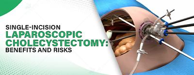
Single-Incision Laparoscopic Cholecystectomy is a minimally invasive gallbladder removal surgery done through one cut, offering quicker recovery and minimal scarring, with some surgical risks.

Expert gastroenterology care for better digestive health, offering advanced diagnosis and treatment for GERD, IBS, ulcers, and liver disorders.
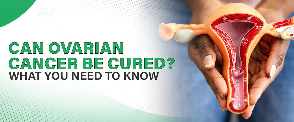
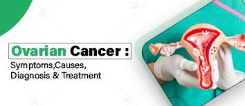
Ovarian cancer is cancer that starts in the ovaries — the female organs that release eggs and hormones such as estrogen and progesterone. It has been referred to as a "silent killer," as the initial symptoms are nonspecific or simply attributed to general maladies such as bloating or pain in the abdomen. This cancer is usually not found until it has already spread within the pelvis and the abdominal cavity, thus, early detection is of utmost importance. Timely follow-up, symptom awareness, and early physician consultation can do a lot to enhcavity ance outcomes.
At the best oncology hospital in Noida, we prioritize awareness and early detection to maximize survival opportunities for women affected by this disease. This guide gives you the vital information regarding ovarian cancer and how it can be well managed.
Call us at +91 9667064100 immediately and book your appointment in Noida for ovarian cancer treatment at the best hospital.
Ovarian cancer, as the name suggests, starts in the ovaries. Ovaries are small, almond-shaped organs that produce eggs and hormones such as estrogen and progesterone. In some cases, ovarian cancer can start in the Fallopian tubes. It has the same symptoms and is treated similarly, even if it comes from the Fallopian tube. No cause has yet been identified, but risk factors can predispose to ovarian cancer.
Signs and symptoms of ovarian cancer are normally nonspecific and may easily be mistaken for routine illnesses. Certain or recurring signs and symptoms, however, should never be ignored and must be assessed by a physician. Some of the described nonspecific red flags are:
Abdominal distension or bloating
Abdominal or pelvic pain
Difficulty in swallowing or rapid fullness
Frequent urge to urinate
Fatigue
Heartburn or indigestion
Changes in bowel movements
Should symptoms continue for more than a few weeks, seek the services from the best doctor for ovarian cancer in Noida for timely assessment and expert care.
There are certain factors that enhance the risk of ovarian cancer:
Age: Ovarian cancer occurs predominantly in women over 60 years of age.
Family History: A familial history of breast or ovarian cancer may raise risk.
Genetic Mutations: Mutations in BRCA1, BRCA2, or Lynch syndrome genes significantly raise the risk.
Reproductive History: Women Women who have never given birth or have given birth at an advanced age might be at greater risk.
Hormone Replacement Therapy: Long-term use of HRT can also increase the chance of developing ovarian cancer.
Obesity and Endometriosis: These conditions can also enhance the risk of ovarian cancer.
Knowledge of these risk factors can aid in prevention and early detection.
Diagnosis of ovarian cancer is a series of steps:
Pelvic Exam: An examination of the abdomen to identify any abnormality.
Imaging Tests: Ultrasound, CT scan, or MRI to examine the ovaries.
Blood Tests: Screening for the level of CA-125, a protein marker that exists in ovarian cancer.
Biopsy: Removal of tissue to test for cancer cells.
Genetic Testing: To determine inherited mutations that increase the risk.
Proper and timely diagnosis will guarantee effective planning of treatment.
Treatment for ovarian cancer depends on the stage and nature of cancer, and on the patient's overall health. Standard treatment modalities are:
Surgery: Initial treatment, with an intent to remove as much of the tumor as possible.
Total Abdominal Hysterectomy: Removal of uterus, ovaries, fallopian tubes, and the lymph nodes in the region.
Debulking Surgery: Removes as much observable tumor as possible, usually followed by chemotherapy.
Chemotherapy: Chemotherapy destroys cancer cells or prevents cancer cell growth with medication.
Intravenous Chemotherapy: Given intravenously into a vein.
Intraperitoneal Chemotherapy: Given within the abdominal space for more localized exposure.
Targeted Therapy: This therapy focuses on targeting very specific molecules that assist cancer cell growth.
PARP Inhibitors: Effective for cancer with BRCA Mutation.
Angiogenesis Inhibitors: Prevents growth of blood vessels feeding tumors.
Hormone Therapy: Blocks hormones from nourishing some forms of ovarian cancer.
Estrogen Receptor Modulators: Prevents estrogen from stimulating cancer cell growth.
Radiation Therapy: Uses high-energy beams to kill cancer cells. Although ovarian cancer patients rarely use this therapy, severe cases may incorporate it into their treatment plan.
Ovarian cancer is managed best by the team of gynecologic oncologists, surgeons, medical oncologists and other specialists who provide holistic care.
Although preventing ovarian cancer is not guaranteed, certain measures can help reduce the risk:
Oral Contraceptives: Prolonged use may reduce risk.
Pregnancy and Breastfeeding: Both are associated with a reduced risk.
Genetic Counselling: For those having a family history or known genetic mutations.
Regular Check-ups: Periodic gynecological check-ups for early identification.
Discussion about individual risk factors with a health care provider can help in decision-making about prevention and screening of ovarian cancer.
If you or someone you know is showing signs of ovarian cancer, don't delay.
Ovarian cancer is a malignant but treatable disease, particularly if it is diagnosed early and with the proper balance of experience and attention. Although a diagnosis may be frightening, advances in surgery, chemotherapy, and biologic therapy are raising hopes and improving survival rates for women. If you or a woman you know is at risk or has symptoms, don't delay seeking professional advice.
At our cutting-edge center in Noida, our gynecologic and oncology specialists strive to deliver personalized care with compassion and accuracy. Schedule a consultation today to regain control over your health at an affordable cost in Noida.
Q1. Is it possible for ovarian cancer to occur even after menopause?
Ans: Yes. Ovarian cancer is most commonly diagnosed in women over the age of 60 years, and again, most of them are postmenopausal. While hormone levels fall after menopause, with increasing age, the risk for ovarian cancer grows, so check-ups are essential.
Q2. If my CA-125 is normal, can I still have ovarian cancer?
Ans: Yes. Although CA-125 is a useful marker, it does not necessarily increase in early ovarian cancer. There are some women with cancer who have normal CA-125, and there are some who have increased CA-125 without cancer. For this reason, doctors perform the test in conjunction with imaging and other procedures.
Q3. In what way does ovarian cancer impact fertility, and are options available to preserve it?
Ans: Treatment for ovarian cancer and its impact on fertility—surgery and chemotherapy, for instance—can have effects on fertility. However, for young women diagnosed with ovarian cancer at an early stage, options may include cryopreservation of eggs or embryos before treatment and fertility-sparing procedures.
Q4. Is chemotherapy always required for the treatment of ovarian cancer?
Ans: No. Chemotherapy depends on stage, grade, and tumor type. In extremely early-stage or low-grade tumors, surgery alone might be enough. Your oncology team will determine the optimum approach based on a complete pathology report.
Q5. Can lifestyle factors affect ovarian cancer recurrence risk?
Ans: Yes. Although no change in lifestyle can promise prevention, maintaining a healthy weight, a nutritious diet, exercise, and no smoking potentially lower the risk for recurrence and overall health during recovery.
Q6. Are the symptoms of ovarian cancer different from those of usual disorders such as PCOS or IBS?
Ans: Yes. Although there is a degree of overlap in symptoms such as bloating or pain in the pelvis, ovarian cancer symptoms tend to be more persistent and progressive. Unlike PCOS or IBS, ovarian cancer symptoms typically do not fluctuate with menstrual cycles or dietary changes.

The gallbladder may be tiny, but it plays an important role in digesting your food. Hidden beneath your liver, this pear-shaped organ is responsible for storing bile, a liquid that assists in breaking down fat in your food. Pain or even serious health issues result when things go wrong, like when gallstones form. At the top gallbladder hospital in Greater Noida, we are here to guide you to catch the red flags early. Knowledge of symptoms of gallbladder stones is important, so let us explore what to do and why swift action is essential.
Gallstones do not always yell—but when they yell, we hear. Contact the top hospitals for early detection and customized treatment before symptoms worsen. Call us now at +(91) 9667064100 for expert consultation.
Gallstones are hard, pebble-sized growths that form in the gallbladder when the chemicals in bile, such as cholesterol or bilirubin, become imbalanced. They can be smaller than a grain of sand or larger than a golf ball. Some do nothing but harm, but others clog bile ducts, causing pain and trouble. Gallbladder problems don't remain localized—these can travel to adjacent organs such as the liver or pancreas if left untreated. Catching symptoms early is key to avoiding bigger problems.
Several factors raise your risk of gallstones:
Diet: Consuming excessive amounts of fatty or processed food can alter the balance, leading to the formation of stones.
Weight: Being overweight or losing weight too fast ups the odds.
Genetics: If your family has a history, you’re more likely to get them.
Health Conditions: Diabetes or liver issues can mess with bile production.
Lifestyle: Fasting or skipping meals may slow down bile flow, leading to a buildup.
In the best hospitals, they diagnosed gallstones in every age group, but they occur more in women, individuals above 40, and those with inactive lifestyles. Being aware of the causes is helpful, but identifying symptoms leads to getting help quickly.
Gallstones are not always problematic, but when they do cause problems, the symptoms won't be easy to ignore. Here's a close-up view of the symptoms you shouldn't ignore:
The traditional gallbladder symptom is pain on your upper right side, just below your ribcage. It may be a dull ache, a tight cramp, or a stabbing feeling. It usually hits after you eat heavy or greasy food, such as butter chicken or fried snacks, as the gallbladder finds it difficult to release bile. Pain may last for minutes or hours, sometimes waking you up at night. If the pain is severe or persistent, do not wait—this is a red flag.
Feeling queasy after meals, especially greasy ones, is common with gallstones. When bile can’t flow properly, digestion falters, leading to nausea or even vomiting. This isn’t just a bad stomach day—if nausea hits regularly with belly pain, your gallbladder might be signaling trouble.
Ever feel bloated, gassy, or too full after meals? Gallstones may hinder bile flow, and it becomes difficult to digest fat. This results in indigestion, belching, or a full stomach. If accompanied by pain or nausea, it's time for your gallbladder to be checked. Try a food diary to catch patterns—bring it up to your doctor for a better diagnosis.
A temperature with a stomachache isn't usual—it may be a sign of inflammation (cholecystitis) or infection from an obstructed bile duct. Chills may accompany this, so you might feel cold and ill. This combination requires immediate attention, since infections that go untreated can develop quickly into serious complications.
If your eyes or skin become yellow, it's a sign. Gallstones may cause bile ducts to become blocked, and when that happens, bilirubin (a pigment in bile) builds up in your blood. This jaundice is usually accompanied by itchy skin or tiredness. It's not only for show—it alerts you to a blockage that may require surgical correction. Don't wait to seek a specialist if this is the case.
Bile also colors your stool brown, so a blocked duct may cause stools to appear clay-colored or pale. Excess bilirubin, on the other hand, darkens urine to a deep amber color. These symptoms in combination are shouting gallbladder trouble—get them checked ASAP.
Gallstone pain does not always remain stationary. It may refer to your right shoulder blade or upper back as a muscle ache or knot. This "referred pain" occurs due to nerve pathways. If pain in the belly radiates like this, particularly after eating, your gallbladder could be the problem.
Small steps can reduce your risk:
Eat Regularly: Missed meals disrupt bile flow—maintain regular mealtimes.
Lose weight gradually: 1-2 pounds per week prevents stone development.
Select Fiber: Fruits, vegetables, and whole grains keep bile in check.
Exercise regularly: Regular exercise maintains a healthy weight and digestion.
Are you still unsure if it's simply gas or something more serious? Let's determine it and go to the best hospital in Noida.
Gallstones may creep up on you, but their symptoms—jaundice, nausea, and pain—cry out. Don't let them take your peace away. With the finest hospital's assistance, you can overcome gallstone problems head-on and return to living life in abundance. Early detection of these signs may spare you pain and concern—reserve an appointment today and prioritize your health!
Q1. Will gallbladder stones clear up by themselves?
Ans: Some small gallstones may not cause symptoms and could remain unnoticed for years. However, once they start blocking bile ducts or causing discomfort, they typically require medical attention. Stones rarely "go away" on their own once symptoms appear.
Q2. After eating, what causes a gallbladder attack?
Ans: Fatty or greasy food tends to precipitate gallbladder attacks. This is due to the contraction of the gallbladder to empty bile, and if a stone in the duct obstructs it, pain results, which is typically in the upper right abdomen or back. High-fat meals, such as fried foods or creamy meals, are frequent offenders.
Q3. Do stones always require removal of the gallbladder?
Ans: Not necessarily. If gallstones are not symptomatic, treatment may not be necessary right away. But if there is pain, inflammation, or obstruction, removal (usually by laparoscopic surgery) is the most effective and common therapy.
Q4. Are there symptoms other than abdominal pain caused by gallstones?
Ans: Yes. Gallstones may produce nausea, vomiting, bloating, indigestion, shoulder or back pain, and even fever or jaundice if there are any complications. These are very common symptoms of other digestive problems as well, so proper diagnosis is essential.
Q5. How are gallstones diagnosed?
Ans: Ultrasound is the most frequently used and reliable method to diagnose gallstones. Other tests, such as liver function tests, HIDA scans, or MRCPs, are done to identify blockage, infection, or associated complications.
Q6. Are untreated gallstones dangerous?
Ans: Although some gallstones are asymptomatic, symptomatic gallstones, if left untreated, can result in severe conditions such as cholecystitis (inflammation), pancreatitis, or infection of the bile ducts. In their worst forms, they may need emergency treatment.
Q7. Will lifestyle modifications actually prevent gallstones?
Ans: Yes. Being at a healthy weight, having balanced meals rich in fiber, avoiding quick weight loss, and exercising regularly can lower your risk of getting gallstones. Eating regular meals and drinking plenty of water also keep bile flowing normally.

Gallstones (Cholelithiasis) are hardened deposits in the gallbladder that cause pain, nausea, and bloating. They are diagnosed via ultrasound and treated with medication or surgery. Call +91 9667064100 for expert care.

Subscribe to our
© Copyright 2026. All Rights Reserved by Felix Healthcare Private Limited
Design and development by :


 +(91)9667064100
 Emergency : +(91)9667064100