 WhatsApp
 WhatsApp


Subscribe to our

Neurological disorders can significantly affect an individual’s daily life, as they arise from abnormalities in the brain, spinal cord or the peripheral nerves. When these vital structures are affected, a person may experience weakness, numbness, seizures, pain, or difficulty with movement, speech or coordination. To properly manage these conditions, it is crucial to understand what are neurological disorders, the types of neurological disorders, and how they are identified and treated. Individuals experiencing the early signs of neurological problems should seek assessment at a qualified top neuro hospital in Noida to prevent complications and ensure timely treatment.
Neurological disorders refer to diseases that affect any part of the nervous system. These may be due to genetic factors, infections, immune reactions, injuries, or degenerative changes. When the nervous system becomes compromised, the person may experience issues ranging from mild discomfort to severe disability. Understanding the causes of neurological disorders allows patients and caregivers to seek medical attention early and improve treatment outcomes.
Awareness of the types of neurological disorders helps patients recognize symptoms early and seek the right care.
Parkinson’s disease is a progressive condition caused by the deterioration of dopamine-producing neurons. This leads to slow movements, tremors, stiffness and balance difficulties. As the disease advances, daily tasks become increasingly challenging, and ongoing treatment is required to manage symptoms.
Alzheimer’s disease is the most common cause of dementia, leading to memory loss and declining cognitive function. Abnormal protein deposits disrupt communication between brain cells, causing behavioral changes and difficulties in performing everyday tasks.
MS is an autoimmune condition where the body attacks its own nerve coverings (myelin). This disrupts nerve signals and leads to weakness, vision issues, imbalance, or fatigue. MS usually follows a pattern of remission and relapse, making long-term monitoring essential.
A stroke occurs when blood flow to the brain is blocked or a blood vessel bursts. Lack of oxygen causes permanent damage to brain tissues, resulting in paralysis, speech problems, confusion or loss of balance. Immediate treatment is critical for a better outcome.
Epilepsy involves recurrent seizures caused by abnormal electrical activity in the brain. Symptoms depend on the type of seizure and may include shaking, staring spells or sudden loss of awareness. Timely treatment helps control seizures and improves quality of life.
Recognizing the symptoms of common neurological diseases can help with early diagnosis and management. These may include:
Recurrent headaches or migraines
Persistent numbness or tingling
Difficulty walking or balancing
Tremors or involuntary movements
Memory loss or confusion
Speech or swallowing problems
Seizures
Chronic back or neck pain
If these symptoms persist, it is advisable to seek a neurologist’s opinion.
The process of diagnosing neurological conditions involves a combination of:
Neurological examination – Assessing reflexes, strength, sensation and balance
Imaging tests – MRI and CT scans to view structural changes
EEG – detecting abnormal electrical activity for epilepsy
EMG/Nerve conduction studies – assessing nerve and muscle function
Lumbar puncture – examining spinal fluid for infections or autoimmune diseases
Accurate diagnosis allows neurologists to form a personalized plan based on the patient’s needs.
At a well-equipped Neurology hospital in Noida, treatment combines medicines, therapies and surgical interventions depending on the condition and severity.
Anticonvulsants for epilepsy
Dopamine-enhancing medicines for Parkinson’s disease
Immunomodulators and steroids for multiple sclerosis
Blood thinners after a stroke
These medicines help control symptoms, prevent disease progression and reduce complications.
Physiotherapy for mobility and balance
Occupational therapy to restore daily functioning
Speech therapy for communication and swallowing issues
Cognitive therapy for memory and behavioral changes
These therapies promote recovery and support long-term independence.
Certain neurological disorders require surgical intervention, especially when symptoms do not respond to medications.
DBS is commonly used for Parkinson’s disease, essential tremors and dystonia. Electrodes are implanted in specific brain regions to regulate abnormal signals and improve mobility.
Surgery is performed to remove benign or malignant brain tumours, relieve pressure, restore brain function and prevent further neurological decline.
For patients with brain aneurysms, surgical clipping or minimally invasive coiling is done to prevent bleeding and life-threatening complications.
Endovascular procedures can remove clots from blocked arteries, improving recovery chances when performed within the critical time window.
For spinal cord compression, slipped discs or nerve compression, surgical decompression may be required to relieve symptoms like pain and weakness.
These techniques improve outcomes significantly when performed by experienced neurosurgeons.
People should seek medical attention if they notice:
Sudden weakness on one side of the body
Persistent headaches
Repeated episodes of dizziness
Memory loss affecting daily life
Seizures or fainting spells
Difficulty speaking or understanding
Changes in behaviour or personality
Visiting a neurologist early ensures timely diagnosis and better treatment outcomes.
Understanding what neurological disorders are, their symptoms, and the available treatment approaches helps individuals take control of their health. Visiting a well-equipped Neuro hospital in Noida or consulting the Best Neurologist in Noida ensures timely diagnosis, advanced treatment options, and ongoing support for long-term management. Whether symptoms are mild or severe, early intervention can significantly improve recovery, mobility, and overall quality of life. Anyone noticing the early signs of neurological problems should not delay evaluation, as prompt care plays a vital role in maintaining neurological health.
Q. 1. When should I worry about persistent headaches?
Ans. If headaches last for weeks or affect daily activities, a neurological evaluation is recommended.
Q. 2. Can neurological disorders be cured permanently?
Ans. Some conditions can be cured, while others can be effectively managed with long-term treatment.
Q. 3. Are all seizures a sign of epilepsy?
Ans. No, seizures can also occur due to fever, infections or metabolic issues and need proper diagnosis.
Q. 4. Is surgery always required for neurological disorders?
Ans. No, surgery is suggested only when medicines and therapies are not sufficient.
Q. 5. How can I identify early signs of neurological problems in children or adults?
Ans. Look for changes in movement, speech, behaviour, memory or unexplained weakness.
Advanced neurosurgery improves treatment for brain and spine disorders with better outcomes.
Experiencing tingling in your hands or feet? It could signal a nerve-related issue. Consult our expert neurologists at Felix Hospitals to find the cause and get the right treatment today.
Neurological disorders impact the brain, spine, and nerves, causing symptoms like headaches, seizures, or memory loss. Discover causes, treatments, and expert advice for better neurological care and management.

Presbyopia, commonly referred to as age-related farsightedness, is a widespread vision problem that affects millions globally as they grow older. This condition makes it difficult to focus on nearby objects, impacting everyday activities such as reading, sewing, or using a smartphone. It arises due to natural eye aging and changes in the lens, resulting in diminished flexibility. Families and individuals seeking specialized care can rely on a presbyopia hospital in Noida, where experienced professionals offer advanced treatments aimed at maintaining eye health and improving quality of life.
In this detailed blog, readers will learn about presbyopia, related vision problems, its symptoms, underlying causes such as lens changes, and explore effective presbyopia treatment methods along with preventive strategies. Armed with this knowledge, patients can make informed decisions about when to consult an eye specialist in Noida for timely vision correction.
Call +91 96670 64100 now to start your journey toward better vision and eye comfort.
Presbyopia is a vision condition caused by the gradual loss of the eye lens’s ability to focus on nearby objects, leading to significant near vision difficulty. This condition usually emerges after age 40 and worsens progressively as the lens inside the eye stiffens and loses its elasticity. Unlike other typical refractive errors that depend on the shape of the eyeball, presbyopia results from natural lens changes due to aging.
In youth, the eye’s lens is flexible and can easily change shape to focus light on the retina clearly at various distances, a process called accommodation. With advancing age, the lens hardens and loses this ability to adjust, making close objects appear blurry. Activities that were once effortless, such as reading small print or threading a needle, become challenging.
The symptoms of presbyopia usually develop gradually and manifest as:
A need to hold reading materials at arm’s length or farther away to see clearly
Blurred vision while doing close-up activities like reading or writing
Eyestrain or fatigue after prolonged near-vision tasks
Frequent headaches linked to eye strain
A requirement for brighter light when reading
Difficulty in seeing and focusing in dim lighting conditions
Squinting to bring close objects into focus
If left uncorrected, these symptoms worsen, causing challenges in daily life. Early diagnosis at a presbyopia hospital in Noida can help manage these symptoms effectively, improving vision and comfort.
The primary cause of presbyopia is the gradual stiffening of the eye’s lens due to natural eye aging. Over time, the proteins in the lens change, resulting in thickening and reduced flexibility. This stiffening compromises the lens's ability to change shape and focus on nearby objects. Risk factors contributing to presbyopia include:
Age over 40 years, the most important factor
Pre-existing medical conditions like diabetes, which affect eye function
Previous eye surgeries or injuries
Genetics, which may predispose some individuals to earlier onset
Use of certain medications that can impact focusing ability
Understanding these causes is vital for recognizing early signs and seeking timely care.
By impairing near vision, presbyopia interferes with work, leisure, and self-care tasks, lowering quality of life. Uncorrected presbyopia causes persistent squinting and eye strain, which may lead to fatigue and headaches. Often, presbyopia coexists with other vision problems like nearsightedness (myopia) or farsightedness (hyperopia), complicating corrective measures.
Early treatment is essential to preserve eye health and functionality, helping prevent secondary complications like chronic headaches and eye discomfort.
While presbyopia cannot be reversed, various presbyopia treatment options help restore clear near vision:
These are the most common corrective tools, allowing refocusing of light on the retina during close tasks. Reading glasses are affordable and provide quick relief but must be updated regularly as the condition progresses.
Multifocal or monovision contact lenses offer a noticeable alternative to glasses by providing distinct vision zones for distance and near. These options suit active individuals desiring a glasses-free lifestyle.
Advanced methods designed to improve focusing ability include:
Monovision LASIK, which corrects one eye for distance vision and the other for near tasks
Conductive keratoplasty, which reshapes the cornea using radiofrequency energy
Lens replacement surgery, where the stiff lens is replaced with an artificial intraocular lens to restore accommodation
Surgical options require consultation with an experienced eye specialist in Noida to determine suitability and expectations.
Simple measures such as ensuring adequate lighting, taking breaks during near work, and using magnifying tools help reduce eye strain and ease symptoms.
Though presbyopia is a natural aging process, maintaining overall eye health slows symptom progression and enhances comfort:
Regular comprehensive eye examinations promote early detection and timely prescription updates
A balanced diet rich in antioxidants supports eye tissues
Using UV-protective sunglasses shields eyes from harmful rays
Managing systemic conditions like diabetes and hypertension reduces ocular damage
Avoidance of smoking and excessive alcohol consumption protects vision
Staying hydrated and performing basic eye exercises preserve eye function longer
Visiting a presbyopia hospital in Noida helps provide personalized preventive guidance.
If you experience difficulty focusing on nearby objects or increased eye strain, promptly scheduling a professional eye exam is vital. Early vision correction improves outcomes and prevents lifestyle disruption.
Call +91 96670 64100 for consultations with skilled professionals and comprehensive presbyopia treatment options.
In conclusion, presbyopia is an inevitable, natural part of aging characterized by the gradual loss of the eye’s ability to focus on nearby objects due to lens changes and eye aging. It manifests as near vision difficulty and other vision problems that can significantly affect daily activities such as reading and using smartphones. Fortunately, presbyopia treatment options such as reading glasses, contact lenses, and advanced surgical procedures greatly improve vision clarity and quality of life.
Families and individuals seeking expert care can rely on a presbyopia hospital in Noida, ensuring access to comprehensive diagnosis, personalized treatment plans, and ongoing support from experienced eye specialists in Noida. Early evaluation helps preserve eye health, prevent complications, and enables patients to maintain an active, fulfilling lifestyle.
Q.1. At what age does presbyopia usually start?
Ans. Typically around 40 years old.
Q.2. Can presbyopia be prevented?
Ans. It isn’t preventable but symptoms can be managed and progression slowed.
Q.3 Are reading glasses necessary?
Ans. Yes, they are the simplest correction method.
Q.4. Is presbyopia curable with surgery?
Ans. Surgery improves near vision but isn’t a cure and requires consultation.
Q.5. Does presbyopia affect other vision issues?
Ans. It can coexist with nearsightedness or farsightedness.

Eye flu, also known as conjunctivitis, is a common eye infection causing redness, irritation, and watering. Learn its causes, symptoms, and effective treatments.
The right ophthalmologist is a crucial step in your eye surgery journey
जो लोग लंबे समय से जोड़ों के दर्द, सूजन या अकड़न से परेशान हैं और दवा, फिजियोथैरेपी या अन्य उपायों से राहत नहीं मिल पा रही। उनके लिए जॉइंट रिप्लेसमेंट सर्जरी एक प्रभावी समाधान बनकर सामने आती है। यह सर्जरी सिर्फ बुजुर्गों के लिए नहीं, बल्कि युवा मरीजों के लिए भी उम्मीद की किरण है। खासकर तब, जब जोड़ों का दर्द चलने, उठने-बैठने या काम करने की क्षमता पर असर डालने लगे। Joint Replacement Surgery in Noida में उपलब्ध है। आइए जानें कि जॉइंट रिप्लेसमेंट सर्जरी से पहले क्या तैयारी करनी चाहिए। इसकी लागत कितनी होती है और नोएडा में बेहतरीन इलाज कहां मिलता है।
अभी अपॉइंटमेंट शेड्यूल करें – कॉल करें: +91 9667064100
जॉइंट रिप्लेसमेंट सर्जरी एक आर्थोपेडिक प्रक्रिया है। जिसमें खराब या घिसे हुए जोड़ों जैसे घुटना, हिप या कंधा को कृत्रिम इम्प्लांट से बदलाता है। इस सर्जरी का उद्देश्य दर्द को कम करना, जोड़ों की गतिशीलता वापस लाना और जीवन की गुणवत्ता में सुधार करना है। यह सर्जरी ऑस्टियोआर्थराइटिस या रुमेटॉइड आर्थराइटिस, हड्डी का फ्रैक्चर (Bone fracture) या चोट, जन्मजात विकार, अवस्कुलर नेक्रोसिस व जोड़ों में कैल्सिफिकेशन या विकृति के इलाज के लिए होती है।
डॉक्टर से विस्तृत परामर्श लें:
अपने आर्थोपेडिक सर्जन से सर्जरी के लाभ, संभावित जोखिम, इम्प्लांट के प्रकार और रिकवरी की प्रक्रिया के बारे में विस्तार से जानें।
मेडिकल हिस्ट्री बताएं:
अगर आपको ब्लड प्रेशर, डायबिटीज, हार्ट या लंग्स की समस्या है तो डॉक्टर को जरूर बताएं। सर्जरी से पहले इनका नियंत्रण जरूरी है।
दवाओं की जानकारी दें:
अगर आप ब्लड थिनर या स्टेरॉयड ले रहे हैं तो सर्जन को सूचित करें, क्योंकि सर्जरी से पहले इन्हें अस्थायी रूप से रोकने की सलाह दी जाती है।
सर्जरी से पूर्व मरीज की पूरी मेडिकल फिटनेस जांच की जाती है ताकि ऑपरेशन के दौरान किसी प्रकार की जटिलता न हो और रिकवरी प्रक्रिया सुचारु रूप से हो सके। इसके अंतर्गत निम्न प्रमुख जांचें शामिल हैं।
ब्लड टेस्ट:
इससे मरीज के हीमोग्लोबिन, ब्लड शुगर, किडनी और लिवर फंक्शन की जानकारी मिलती है इंफेक्शन, ब्लीडिंग टेंडेंसी या एनीमिया जैसी स्थितियों का पता लगाया जाता है।
यूरिन टेस्ट:
मूत्र में संक्रमण (यूटीआई) या किडनी से जुड़ी समस्या की पहचान की जाती है। ऑपरेशन के बाद संक्रमण की संभावना कम करने में यह टेस्ट मददगार होता है।
ईसीजीः
दिल की विद्युत गतिविधियों की जांच के लिए किया जाता है। इससे यह सुनिश्चित किया जाता है कि सर्जरी से पहले मरीज का हृदय पूरी तरह स्वस्थ है।
एक्स-रे:
फेफड़ों की स्थिति और छाती के अंदरूनी हिस्सों की स्थिति का पता लगाया जाता है। इससे सर्जरी के दौरान एनेस्थीसिया देने में मदद मिलती है।
एमआरआई या सीटी स्कैनः
यदि सर्जरी हड्डियों, जोड़ों, दिमाग या रीढ़ से संबंधित है तो एमआरआई या सीटी स्कैन जरूरी होता है। इससे डॉक्टर को सटीक लोकेशन और जटिलताओं की पूरी जानकारी मिलती है।
कार्डियोलॉजिस्ट या फिजिशियन से क्लीयरेंस:
सर्जरी से पहले विशेषज्ञ डॉक्टर (कार्डियोलॉजिस्ट/फिजिशियन) मरीज की सभी रिपोर्ट देखकर फिटनेस सर्टिफिकेट जारी करते हैं। यदि मरीज को पहले से कोई बीमारी (जैसे डायबिटीज, ब्लड प्रेशर या अस्थमा) है, तो उसे कंट्रोल में लाने के निर्देश दिए जाते हैं।
फेफड़ों और किडनी की अतिरिक्त जांच:
लंबे समय से बीमार मरीजों या बुजुर्गों के लिए फेफड़े और किडनी की जांच भी की जाती है। यह सुनिश्चित किया जाता है कि शरीर की ऑक्सीजन और विषाक्त पदार्थ निकालने की क्षमता सही ढंग से काम कर रही हो।
एलर्जी और दवा प्रतिक्रिया जांच:
यह जांच पता लगाती है कि मरीज को किसी दवा या एनेस्थीसिया से एलर्जी तो नहीं है। इससे ऑपरेशन के दौरान दवा से जुड़ी कोई प्रतिक्रिया होने का खतरा घटता है।
संपूर्ण स्वास्थ्य मूल्यांकन:
वजन, ब्लड प्रेशर, पल्स और तापमान की नियमित जांच की जाती है। ऑपरेशन से पहले डॉक्टर मरीज की मानसिक और शारीरिक स्थिति का भी आकलन करते हैं।
नोएडा में जॉइंट रिप्लेसमेंट कराने के लिए एक मान्यता प्राप्त, अनुभवी डॉक्टर और आधुनिक सुविधाओं वाले अस्पताल का चुनाव बेहद जरूरी है। Complete Guide for Preparing for Best hospital and surgeon उपलब्ध है।
हॉस्पिटल चुनते समय ध्यान दें:
एनएबीएच या आईएसओ प्रमाणित हॉस्पिटल
अनुभवी ऑर्थोपेडिक सर्जन
न्यूनतम इंफेक्शन रेट
अत्याधुनिक मॉड्यूलर ऑपरेशन थिएटर
पोस्ट-सर्जरी रिहैबिलिटेशन और फिजियोथेरेपी (Physiotherapy) यूनिट
लागत कई कारकों पर निर्भर करती हैः
| प्रकार | औसत लागत (रुपये में) | विवरण |
| घुटना रिप्लेसमेंस | 1.8–4.5 लाख | सिंगल नी रिप्लेसमेंट |
| हिप रिप्लेसमेंस | 2–6 लाख | यूनिलेटरल या बाइलेटरल |
| शोल्डर रिप्लेसमेंस | 2.5–5 लाख | टोटल/पार्शियल |
| सरकारी हॉस्पिटल | 1–2 लाख | सीमित सुविधा पर कम लागत |
| निजी हॉस्पिटल | 2.5 – 6 लाख | बेहतर इम्प्लांट और तकनीक |
आयुष्मान भारत, सीजीएचएस, ईसीएचएस और कई निजी बीमा पॉलिसियां जॉइंट रिप्लेसमेंट को कवर करती हैं। इससे मरीज का खर्च 60–90% तक कम हो सकता है।
सर्जरी से पहले और बाद में फिजियोथैरेपी बेहद जरूरी है। सर्जरी से पहले प्री-हैब एक्सरसाइज से मांसपेशियां मजबूत बनाएं। सर्जरी के बाद धीरे-धीरे मूवमेंट, बैलेंस और वॉकिंग ट्रेनिंग करें। मानसिक रूप से खुद को तैयार रखें कि शुरुआती दिनों में थोड़ी असुविधा होगी, लेकिन नियमित अभ्यास से जीवन सामान्य हो जाएगा।
छोटी-सी लापरवाही भी संक्रमण, सूजन या दोबारा दर्द जैसी समस्याएँ पैदा करती है। इसलिए मरीज को विशेष ध्यान देना चाहिए।
एक्सरसाइज रूटीन का पालन करें:
सर्जरी के बाद शरीर की गतिशीलता को वापस लाने के लिए फिजियोथैरेपी बहुत जरूरी है। एक्सरसाइज से मांसपेशियों में जकड़न नहीं होती और रक्त संचार सामान्य रहता है। डॉक्टर या फिजियोथैरेपिस्ट (Physiotherapist) की सलाह के बिना किसी नई एक्सरसाइज की शुरुआत न करें।
जमीन पर बैठने या ज्यादा झुकने से बचें:
विशेषकर जोड़ों या रीढ़ की सर्जरी के बाद, नीचे बैठना या आगे झुकना जोड़ों पर दबाव डालता है। इससे स्टिच खुलने, दर्द बढ़ने या अंदरूनी चोट के खतरे बढ़ जाते हैं। कुर्सी या ऊंचे स्थान पर बैठने की आदत डालें और झुकने वाले कार्यों से परहेज करें।
भारी वजन उठाने से परहेज़ करें:
सर्जरी के बाद कम से कम 2–3 महीने तक भारी वस्तुएं उठाना सख्त मना होता है। इससे सर्जरी वाले हिस्से पर दबाव पड़ सकता है और हीलिंग प्रक्रिया धीमी हो सकती है। रोजमर्रा के कार्यों में भी शरीर पर अतिरिक्त तनाव डालने से बचें।
ऑपरेशन साइट की सफाई और देखभाल:
ऑपरेशन वाले हिस्से को हमेशा साफ और सूखा रखें। पट्टियां (ड्रेसिंग) डॉक्टर की सलाह के अनुसार समय पर बदलें। किसी भी प्रकार की लाली, सूजन, मवाद या बदबू महसूस हो तो तुरंत डॉक्टर को सूचित करें। ये संक्रमण के संकेत होते हैं।
दवाओं का सेवन डॉक्टर की सलाह से करें:
एंटीबायोटिक और पेनकिलर केवल डॉक्टर द्वारा बताए गए समय और डोज़ में ही लें। बिना परामर्श के कोई दवा बदलना या छोड़ना सही नहीं है। पर्याप्त पानी पिएं ताकि दवाओं का असर बेहतर हो और शरीर में टॉक्सिन्स न जमा हों।
नियमित फॉलोअप विजिट करें:
हर 3 से 6 महीने में डॉक्टर से जांच करवाना जरूरी है, ताकि रिकवरी की प्रगति का मूल्यांकन हो सके। एक्स-रे, एमआरआई या ब्लड टेस्ट जैसी जांचें डॉक्टर की सलाह पर दोबारा कराई जा सकती हैं। अगर दर्द, सूजन या चलने-फिरने में परेशानी महसूस हो तो अगली विजिट से पहले ही डॉक्टर से संपर्क करें।
संतुलित आहार और पर्याप्त आराम:
प्रोटीन, विटामिन और मिनरल्स से भरपूर भोजन लें ताकि घाव जल्दी भरें। नींद पूरी करें और तनाव से दूर रहें। यह शरीर की रिकवरी प्रक्रिया को तेज करता है। धूम्रपान और शराब से पूरी तरह दूरी बनाएं, क्योंकि ये हीलिंग को धीमा करते हैं।
जॉइंट रिप्लेसमेंट सर्जरी आज एक सुरक्षित और असरदार प्रक्रिया है। जो लाखों लोगों के जीवन में नई गतिशीलता ला रही है। सही तैयारी, अनुभवी डॉक्टर, आधुनिक तकनीक और पोस्ट-सर्जरी फिजियोथैरेपी यही सफलता की कुंजी हैं। यदि आप नोएडा या आसपास रहते हैं, तो अनुभवी ऑर्थोपेडिक सर्जन (Best Orthopedics in Noida) से सलाह लेकर सर्जरी की प्लानिंग करें और दर्द-मुक्त जीवन की शुरुआत करें। इलाज में बिल्कुल भी देरी नहीं करनी चाहिए। ऐसा करना नुकसानदेह हो सकता है।
अभी अपॉइंटमेंट शेड्यूल करें – कॉल करें: +91 9667064100
प्रश्न.1: क्या जॉइंट रिप्लेसमेंट सर्जरी दर्दनाक होती है?
उत्तरः सर्जरी के दौरान एनेस्थीसिया दिया जाता है। जिससे कोई दर्द नहीं होता। बाद में दर्द दवाओं और फिजियोथैरेपी से नियंत्रित किया जा ता है।
प्रश्न 2: सर्जरी के बाद कितने दिन में चलना शुरू कर सकते हैं?
उत्तरः अधिकतर मरीज 2–3 दिन में वॉकर के सहारे चलने लगते हैं। 4–6 सप्ताह में सामान्य गतिविधियां कर सकते हैं।
प्रश्न 3: क्या सर्जरी का असर लंबे समय तक रहता है?
उत्तरः हां, आधुनिक इम्प्लांट 15–20 साल तक टिकते हैं। लेकिन इसे अच्छे अस्पताल के डॉक्टर की सलाह पर लगवाना चाहिए।
प्रश्न 4: क्या हिप और नी रिप्लेसमेंट एक साथ कर सकते हैं?
उत्तरः कुछ मामलों में संभव है। लेकिन डॉक्टर की सलाह पर निर्णय लिया जाता है। डॉक्टर की सलाह के बिना कोई भी इलाज नहीं कराना चाहिए।
हिप रिप्लेसमेंट सर्जरी एक सुरक्षित और प्रभावी प्रक्रिया है जो गंभीर जोड़ दर्द से राहत दिलाकर चलने-फिरने की क्षमता को बेहतर बनाती है। इसमें हिप का क्षतिग्रस्त हिस्सा कृत्रिम जोड़ से बदला जाता है।
MRI (Magnetic Resonance Imaging) is an advanced diagnostic test used to detect problems related to the brain, spine, joints, and internal organs.
घुटनों का दर्द उम्र, चोट, या गठिया जैसी समस्याओं के कारण आम हो सकता है। जब दवाएं और फिजियोथेरेपी असर नहीं करतीं, तब घुटने की सर्जरी (जैसे कि घुटने का प्रतिस्थापन) जरूरी हो सकती है।
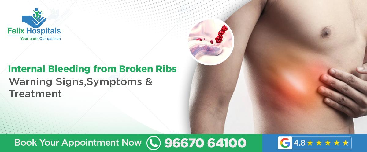
The most typical injuries of falls or accidents or direct traumas to the chest are broken or bruised ribs. Although the pain and discomfort caused by rib fractures may be very high, one of the complications that require to be taken into consideration but may also be quite dangerous is the internal bleeding due to broken ribs. This hemorrhage in the body may cause severe life-threatening diseases that demand immediate attention of a doctor. For those seeking prompt and expert management, locating the right hospital for broken ribs treatment and understanding the factors that influence broken ribs treatment cost is crucial. Consulting the best doctor for broken ribs treatment can ensure correct diagnosis, timely intervention, and a smooth recovery.
Get expert evaluation for broken ribs and related complications at the leading trauma and chest care centers in Noida. Contact +91 96670 64100 .
The ribs safeguard some of the most important organs in the chest such as the lungs, the heart, and great blood vessels. A rib that is displaced or fractured could destroy these underlying organs and blood vessels when broken. The acute, rough edges of fractured or torn ribs may cause a piercing or a tearing of the blood vessels and soft tissues, and cause internal hemorrhage.
Common causes include:
Vehicle crashes, falls, or attacks which have caused severe chest trauma.
Accidents to the chest, sports injuries or high-impact accidents.
With weaker bones, the older adults are even more susceptible to minor trauma effects.
Various fractures of the ribs or flail chest problems will predispose to bleeding.
Comorbidity such as clotting disorders or taking blood thinners that enhance bleeding.
Bruised or broken ribs result in a lot of pain especially when one is breathing deeply, coughing, laughing, or moving the torso. Common symptoms include:
Pain in ribs is sharp or stabbing and is localized to the injured ones.
Chest wall Tenderness, swelling, and occasional bruising.
Pain causes difficulty in deep breathing.
Having a cracking sound when injured.
Loss of mobility due to pains and discomfort.
Most patients who have broken ribs have healed their ribs in weeks, however, the complications such as broken ribs cause internal bleeding, which necessitates urgent medical attention.
Internal bleeding is not very pronounced at the start but may become very sudden. Indications of risk to watch out include:
Sharp chest or abdominal pain which becomes acute.
Breathing problems, dyspnea, or rapid breathing.
Lightheadedness, dizziness or fainting due to a loss of blood.
Lips and extremities turning bluish, which is a sign that they are poorly oxygenated.
Blood pressure and heart rate are low, signs of shock.
Presents blood or blood in sputum, which shows involvement of the lungs.
Anxiety, aching, or pain in the abdomen or chest.
Low blood circulation resulting in paleness or sweating.
The symptoms mentioned are indicative that internal bleeding might be taking place and hence emergency treatment is required in the hospital.
The diagnosis begins with a thorough clinical assessment by the medical practitioner. Physical examination to check bruising, tenderness and listen to the chest using a stethoscope; the doctor will examine the symptoms.
Additional research is:
Chest X-ray: To ensure the number and severity of rib fractures and check lung condition.
CT Scan: Gives in-depth pictures to identify any internal bleeding, injuries to organs or damage of blood vessels.
Ultrasound: to identify the presence of fluid that may suggest bleeding around the lungs or abdomen.
Blood Tests: To examine the level of hemoglobin, coagulation level, and blood loss in general.
Early detection is important to avoid life threatening consequences such as hemothorax or blood buildup in the chest cavity, and the destruction of the lung functions.
The treatment is determined by the severity of bleeding and injury:
Observation and Monitoring: Minor cases of bleeding can be treated through close observation and pain management.
Oxygen Therapy: To make sure that oxygen is delivered adequately in case the lungs are impaired.
Blood Transfusion: And in the case of heavy blood loss so as to stabilize the patient.
Chest Tube Insertion: To remove the blood build up in the chest cavity and avoid collapse of the lungs.
Surgical Intervention: It is necessary in cases of persistent bleeding, huge hematomas, or destruction of organs; it can include fixing or repairing broken ribs, or repairing broken vessels.
Pain Management: Proper analgesia allows the patient to deep breathe and avoid pulmonary infections.
Pulmonary Rehabilitation: Workouts to enhance breathing capacity in the recovery process.
Broken ribs and internal injuries require time to heal but on average six weeks or beyond. Post-discharge imaging and clinical evaluation are to be followed to guarantee that bleeding has stopped and the lung function is restored. Early intervention prevents such complications as pneumonia, chronic pain, respiratory failure.
The overall broken ribs treatment cost depends on:
The severity of injuries and the existence of complications, such as internal bleeding.
Requirement to be admitted, arranged, or hospitalized in need.
Stay and follow-up visits.
Diagnostic imaging and use of blood products.
Rehabilitation and pain management services.
Patients are advised to get detailed cost estimates from the hospital for broken ribs treatment facilities while prioritizing care quality and expertise.
Knowledge on thoracic injuries enhances outcome of patients by delivering:
Proper diagnosis with the help of modern imaging techniques.
Early diagnosis and treatment of intra-operative bleeding.
Expert surgical treatment in case of complicated fractures.
Personalized pain management strategies.
Extensive rehabilitation instructions.
Choosing the best doctor for broken ribs treatment ensures that risks are minimized and recovery is optimized.
Suspect internal bleeding from a rib injury? Seek immediate emergency care by calling + 91 96670 64100.
In summary broken ribs also have internal bleeding that is a serious complication and requires urgent medical treatment. Recognizing warning signs and seeking prompt treatment at a reputed hospital for broken ribs treatment can save lives. Knowledge of the cost of treatment and selection of the appropriate specialist enhances recovery.
Q.1. Do broken ribs result in internal bleeding?
Ans. Yes, sharp pieces of ribs may damage blood vessels, making one bleed internally.
Q.2. How do you know that broken ribs are bleeding?
Ans. Possible internal bleeding is represented by severe chest pain, difficulty breathing, dizziness, and coughing up blood.
Q.3. What are the treatment options of internal bleeding due to broken ribs?
Ans. The available treatment includes monitoring and oxygen therapy to surgery based on the severity of bleeding.
Q.4. What is the time of recovery of fractured ribs?
Ans. Typically 6 weeks, though complications may extend the recovery period.
Q.5. Suffices to diagnose internal bleeding: is the chest X-ray sufficient?
Ans. Chest X-ray detects fractures; CT scan is more sensitive to the detection of internal bleeding.
A pulmonologist plays a crucial role in helping patients dealing with serious or chronic breathing difficulties.
Recognize the types of chest pain that may signal a heart attack and learn about the best treatment options available in Noida for timely care and recovery.

Heart attacks have been identified as one of the major causes of death in the world, and it is important to know when they occur to prevent and treat them better. Interestingly, heart attacks occur more particularly at night and this poses special challenges in detecting and attending to it. If you are concerned about your heart health, consulting a trusted heart specialist in Noida and seeking timely cardiac treatment in Noida can save lives. This blog explores heart attacks at night, night time heart attack causes, heart attack symptoms, signs of heart attack at night, reasons heart attacks are more common at night, and heart attack risk during sleep hours to help you recognize and act promptly.
Schedule a consultation Call on +91 9667064100 with a trusted heart specialist in Noida to receive comprehensive evaluation and personalized cardiac treatment in Noida.
Myocardial infarction or heart attack is a condition that leads to tissue damage or death of a section of the heart muscle due to blocked blood supply. Blockages typically occur following atherosclerosis which is constrained arteries caused by deposition of plaque or abruptly formed clot. Although heart disease may occur at any time of the day, studies show that the occurrence is more common and severe during the night and the early morning hours, which affects patient outcomes.
Several physiological and environmental factors explain why heart attacks at night occur more frequently:
The Circadian Rhythms and Heart Work: The body does have a 24-hour clock, which regulates the amount of hormones released, blood pressure, and the rate of the heart. Cortisol and adrenaline levels change at night influencing blood vessel narrowing and clotting propensity, thus predisposing people to heart attacks.
Blood Pressure Changes during Sleep: Blood pressure normally drops when people sleep but when they wake up or when they experience some sleep problems, the pressure can skyrocket, placing a heart strain and allowing the vessel to rupture.
Changes of the Autonomic Nervous System: Nocturnal alterations in sympathetic and parasympathetic activities result in vascular alterations, platelet aggregation, and inflammation, which may cause heart attacks.
Sleep Apnea and Oxygen Desaturation: Obstructive sleep apnea is typical during the night, and it leads to the frequent decrease of oxygen levels that force the cardiovascular system and risk clot development.
Decreased Symptom Awareness: Awareness of symptoms during sleep or in the early morning may be missed or be mistaken and more damage will be caused.
Recognizing signs of heart attack at night can be lifesaving. The symptoms can marginally vary with daytime symptoms although usually encompass:
Sharp chest pain or uneasiness, which may be termed as pressure or squeezing and fullness.
Radiating pain in arms, neck, jaw or back.
Breathing difficulty, occasionally without chest pains.
Profuse sweating or cold sweat.
Nausea or vomiting
Fatigue and dizziness
Breast pains or irregular heartbeat.
Since the symptoms can wake an individual up, that is why any individual who has such symptoms at night should go to the emergency care.
The causes of heart attacks at night are similar to those that happen during the day, but they also have some unique factors that are related to how the body works at night:
Increased Coagulation Activity: At night, levels of coagulation factors rise, which makes arteries more likely to become blocked.
Changes in vascular tone: Blood vessels get narrower when people sleep, which can lead to ischemia.
Hormonal changes: The peaks of cortisol and noradrenaline affect the risk of heart disease.
Disturbances during sleep: Conditions like sleep apnea cause hypoxia, which increases oxidative stress and inflammation.
These causes lead to awareness that is useful in preventive measures like management of sleep disorders and maximization of medication time.
Studies identify an elevated heart attack risk during sleep hours, particularly between midnight and 6 am, with notable peaks around 3-4 am. Contributing factors are:
Manage high blood pressure or diabetes.
Smoking and alcohol use
Obesity and high cholesterol.
The effects of stress and anxiety on sleep quality.
Abnormal sleeping habits or insomnia.
Patients with these risk factors should prioritize cardiac check-ups and screening with a heart specialist in Noida for tailored risk management and appropriate cardiac treatment in Noida.
To reduce the risk of heart attacks at night, consider the following:
Regular heart examinations: The risks can be identified early and thus addressed in time.
Treat sleep disorders: CPAP or lifestyle changes can treat sleeping disorders such as sleep apnea.
Drug compliance: Take cardiovascular medications regularly, preferably at night to maximize the effect.
Healthy lifestyle: A healthy diet, exercise, quitting smoking, and drinking alcohol moderately reduces risk.
Stress management approaches: Mindfulness, relaxation and proper sleep hygiene are beneficial to the heart.
Emergency preparedness: Know heart attack symptoms and seek immediate help if they occur at night.
If you experience recurrent nighttime chest discomfort, unexplained fatigue, or other suspicious symptoms, consulting a reputable heart specialist in Noida is critical. Early diagnosis and customized cardiac treatment in Noida improve prognosis and reduce complications.
Get a call back now on +(91)96670 64100 and get your heart in expert care.
In conclusion, heart attacks at night are caused by complex physiological changes and have unique risk factors. Recognizing signs of heart attack at night and understanding the night time heart attack causes is crucial for prevention and prompt treatment. Engaging a skilled heart specialist in Noida and accessing quality cardiac treatment in Noida enhances survival and recovery outcomes.
Q.1. What is the reason, why do heart attacks happen during the night?
Ans. Nighttime vessel constriction and clotting is increased by natural body rhythms and hormone variations.
Q.2. Are heart attack symptoms different at night?
Ans. The symptoms are close but can be confused with other disorders or remain undetected when one is sleeping.
Q.3. What can I do to avoid night heart attacks?
Ans. They control risk factors, sleep disorders, taking prescribed medications and healthy lifestyle.
Q.4. Does it make more harm when there is a heart attack at night?
Ans. Delayed symptom recognition and treatment are negative factors that cause more severe damage during night-time heart attacks.
Q.5. When do I need to visit a cardiologist when experiencing a night time pain in the chest?
Ans. Take emergency treatment when the chest pains or symptoms arise, particularly during the night.
In Noida hospitals, recognized for being among the best heart health hospitals in Noida, are at the forefront of offering critical care for heart-related issues.

Find the best cardiologist for your heart care with these essential tips and expert advice.
घुटने की चोट या दर्द केवल खिलाड़ियों तक सीमित नहीं है। अब यह हर उम्र के लोगों में आम है। खासकर जब घुटने में दर्द, सूजन या मूवमेंट में दिक्कत लगातार बनी रहती है, तो आर्थोस्कोपी एक सुरक्षित और प्रभावी समाधान है। Arthroscopic Knee Surgery Treatment in Noida में उपलब्ध है। नोएडा के ऑर्थोपेडिक हॉस्पिटल्स अब Arthroscopic Knee Surgery की अत्याधुनिक सुविधा प्रदान कर रहे हैं। जहां कम चीरे, कम दर्द और जल्दी रिकवरी के साथ इलाज संभव है।
अभी अपॉइंटमेंट शेड्यूल करें – कॉल करें: +91 9667064100
आर्थोस्कोपिक घुटने की सर्जरी (Knee surgery in noida) एक मिनिमली इनवेसिव तकनीक है। जिसमें बड़े चीरे की जगह केवल 2–3 छोटे चीरे लगते हैं। इनसे डॉक्टर एक पतला कैमरा और सूक्ष्म शल्य उपकरण अंदर डालते हैं। कैमरे से घुटने के अंदर का दृश्य मॉनिटर पर देखा जाता है और उसी के आधार पर सर्जरी की जाती है। यह तकनीक एसीएल, पीसीएल, मेनिस्कस, कार्टिलेज या घुटने के अन्य लिगामेंट की चोटों में उपयोग की जाती है।
आर्थोस्कोपिक सर्जरी तब की जाती है जब दवाओं, इंजेक्शन या फिजियोथेरेपी से राहत नहीं मिलती और घुटने की संरचना में वास्तविक क्षति या अस्थिरता होती है। मुख्य स्थितियां जिनमें आर्थोस्कोपिक सर्जरी की जाती है:
एसीएल या पीसीएल लिगामेंट फटना:
घुटने की स्थिरता बनाए रखने वाले मुख्य लिगामेंट्स के फटने पर सर्जरी आवश्यक होती है। खेल गतिविधियों या हादसों में ये चोट आम है। आर्थोस्कोपी द्वारा फटे लिगामेंट का पुनर्निर्माण किया जाता है।
मेनिस्कस का फटना:
मेनिस्कस घुटने के बीच का कुशन होता है जो झटके सोखता है। फटने पर घुटना लॉक होता है। दर्द, सूजन और क्लिकिंग की समस्या होती है। सर्जरी में फटा हिस्सा सिलकर या हटाकर जोड़ को सामान्य किया जाता है।
कार्टिलेज का घिस जाना या टूटना:
जोड़ की हड्डियों के सिरों पर लगी मुलायम परत (कार्टिलेज) के क्षतिग्रस्त होने पर दर्द और रगड़ महसूस होती है। आर्थोस्कोपिक तकनीक से क्षतिग्रस्त हिस्से को समतल या रिपेयर किया जाता है।
जोड़ में ढीलापन या अस्थिरता:
पुराने लिगामेंट या मेनिस्कस चोटों के कारण घुटना कमजोर होता है। आर्थोस्कोपी से टिश्यू को रिपेयर कर स्थिरता बहाल की जाती है।
हड्डी या टिश्यू के टुकड़ों का जोड़ में फंसना:
दुर्घटना या गठिया में हड्डी के छोटे टुकड़े जोड़ के अंदर फंस जाते हैं। इससे मूवमेंट में रुकावट या क्लिक जैसी आवाज आती है। सर्जरी से इन्हें निकालकर जोड़ को मुक्त किया जाता है।
घुटने में लगातार दर्द, सूजन या मूवमेंट की कमी:
लंबे समय से चल रहे आर्थराइटिस (Arthritis), चोट या सूजन में जब अन्य उपचार असफल हों। आर्थोस्कोपी द्वारा जोड़ की सफाई की जाती है जिससे दर्द और सूजन कम होती है।
रोटेशनल या कॉम्प्लेक्स स्पोर्ट्स इंजरी:
फुटबॉल, क्रिकेट, बैडमिंटन या बास्केटबॉल जैसे खेलों में लगी जटिल चोटों के इलाज में भी यह सर्जरी बेहद उपयोगी है।
घुटने में दर्द और सूजन
चलने या मुड़ने पर क्लिकिंग आवाज
घुटने का फिसलना या मुड़ना
मूवमेंट में कठिनाई या जकड़न
खेल या व्यायाम के दौरान अचानक गिरना या घुटना जवाब देना
आर्थोस्कोपिक सर्जरी से पहले सही निदान बेहद जरूरी होता है। जिससे पता चल सके कि जोड़ की समस्या कितनी गंभीर है और किस हिस्से में क्षति हुई है। डॉक्टर विभिन्न क्लिनिकल और इमेजिंग जांचों के माध्यम से सटीक मूल्यांकन करते हैं। मुख्य जांचें जिनसे निदान किया जाता है:
शारीरिक जांच:
डॉक्टर सबसे पहले घुटने की स्थिरता, मूवमेंट और दर्द की तीव्रता को परखते हैं। जोड़ को मोड़ने-सीधा करने, दबाने या खींचने जैसी तकनीकों से यह पता लगाया जाता है कि कौन-सा लिगामेंट, मेनिस्कस या कार्टिलेज प्रभावित है। सूजन, लालिमा, तापमान में वृद्धि या क्लिक जैसी आवाज़ों को भी नोट किया जाता है। इस जांच से प्रारंभिक अंदाजा लगाया जाता है कि समस्या सर्जरी योग्य है या नहीं।
एमआरआई स्कैन:
यह सबसे विश्वसनीय जांच है जिससे लिगामेंट, मेनिस्कस, कार्टिलेज और सॉफ्ट टिश्यू की क्षति का स्पष्ट चित्र मिलता है। एमआरआई से पता चलता है कि चोट आंशिक है या पूर्ण। सूक्ष्म दरारें, सूजन, रक्तस्राव या मांसपेशियों के फटने जैसी स्थितियां भी दिखाई देती हैं। यह डॉक्टर को सर्जरी की आवश्यकता और उसकी योजना तय करने में मदद करता है।
एक्स-रे:
यह जांच हड्डियों के संरेखण, फ्रैक्चर, बोन स्पर, या ऑस्टियोआर्थराइटिस (Osteoarthritis) की स्थिति का पता लगाने के लिए की जाती है। इससे जोड़ों के बीच की दूरी, हड्डियों के घिसाव और जोड़ में कैल्शियम जमाव जैसी समस्याओं का आकलन होता है। एमआरआई से पहले एक्स-रे प्राथमिक स्क्रीनिंग के रूप में किया जाता है ताकि हड्डियों की स्थिति का अंदाजा मिल सके।
अल्ट्रासाउंड:
सॉफ्ट टिश्यू, टेंडन या फ्लूइड की जांच में उपयोगी है। इससे घुटने में तरल पदार्थ जमा है या नहीं, इसका भी पता चलता है।
आर्थोस्कोपी डायग्नोस्टिक टेस्ट:
कुछ मामलों में जब एमआरआई से भी स्पष्ट निदान नहीं मिलता, तो कैमरे की मदद से जोड़ के भीतर देखकर निदान किया जाता है। इसे “डायग्नोस्टिक आर्थोस्कोपी” कहा जाता है, जो छोटी प्रक्रिया होती है और तुरंत सर्जिकल रिपेयर की भी संभावना रहती है।
एनेस्थीसिया देनाः
सर्जरी से पहले मरीज को स्पाइनल या जनरल एनेस्थीसिया दिया जाता है। जिससे उसे कोई दर्द या असुविधा न हो। डॉक्टर मरीज की स्वास्थ्य स्थिति और सर्जरी के प्रकार के अनुसार एनेस्थीसिया का चयन करते हैं। प्रक्रिया के दौरान मरीज पूरी तरह आराम की स्थिति में रहता है।
छोटे चीरे लगानाः
घुटने के आसपास लगभग 2 से 3 छोटे चीरे लगाए जाते हैं। प्रत्येक लगभग 0.5 से 1 सेंटीमीटर के होते हैं। इन चीरे के माध्यम से कैमरा और सर्जिकल उपकरण जोड़ों के भीतर डाले जाते हैं छोटे चीरे होने के कारण घाव जल्दी भर जाते हैं और संक्रमण का खतरा भी बहुत कम रहता है।
कैमरा और उपकरणों का प्रयोगः
एक चीरे से आर्थोस्कोप नामक पतला कैमरा डाला जाता है। जो जोड़ के भीतर की तस्वीर को हाई-डेफिनिशन मॉनिटर पर दिखाता है। दूसरे चीरे से सूक्ष्म सर्जिकल उपकरण जैसे शैवर, कैंची, या ग्रास्पर डाले जाते हैं। इससे डॉक्टर जोड़ की अंदरूनी संरचना को स्पष्ट रूप से देखकर सटीक उपचार करते हैं।
क्षतिग्रस्त टिश्यू की मरम्मत:
कैमरे की मदद से डॉक्टर क्षतिग्रस्त लिगामेंट, मेनिस्कस या कार्टिलेज की पहचान कर उन्हें रिपेयर या हटाते हैं। यदि मेनिस्कस फटा हुआ हो, तो उसे सिलाई (Suturing) के जरिए जोड़ा जाता है। कार्टिलेज डैमेज होने पर खराब हिस्से को हटाकर सतह को स्मूद किया जाता है।
नए लिगामेंट का प्रत्यारोपण:
यदि लिगामेंट पूरी तरह फट चुका है, तो नई ग्राफ्ट लगाई जाती है। जिसे एसीएल या पीसीएल कहा जाता है। यह ग्राफ्ट मरीज के शरीर के किसी अन्य हिस्से (जैसे हैमस्ट्रिंग या पेटेलर टेंडन) से ली जाती है या डोनर से प्राप्त की जाती है। ग्राफ्ट को हड्डी में विशेष स्क्रू या फिक्सेशन डिवाइस से जोड़ा जाता है ताकि यह स्थिर हो सके।
प्रक्रिया की अवधि:
पूरी प्रक्रिया सामान्यतः 60 से 90 मिनट में पूरी हो जाती है।यदि एक से अधिक संरचनाएं रिपेयर की जा रही हों तो समय थोड़ा बढ़ सकता है।
पोस्ट-ऑपरेशन देखभालः
सर्जरी के बाद छोटे चीरे सिल दिए जाते हैं और बैंडेज लगाया जाता है। मरीज को सामान्यतः उसी दिन या अगले दिन डिस्चार्ज कर दिया जाता है। डॉक्टर फिजियोथेरेपी और घुटने को धीरे-धीरे चलाने की सलाह देते हैं ताकि मूवमेंट सामान्य हो सके।
ब्लड टेस्ट, ईसीजी, एक्स-रे और एमआरआई करवाएं।
शुगर, बीपी और वजन नियंत्रित रखें।
डॉक्टर से दवाओं, एलर्जी और पुरानी बीमारियों पर चर्चा करें।
सर्जरी से पहले फिजियोथेरेपी से मांसपेशियों को मजबूत करना लाभदायक होता है।
1–2 दिन अस्पताल में निगरानी में रहना होता है।
दर्द और सूजन के लिए दवा व बर्फ की सिकाई करें।
संक्रमण से बचने के लिए पट्टी को सूखा रखें।
बैसाखी या नी-ब्रेस का प्रयोग डॉक्टर की सलाह से करें।
नियमित फिजियोथेरेपी आवश्यक है। यही रिकवरी की कुंजी है।
आर्थोस्कोपिक सर्जरी पारंपरिक (ओपन) सर्जरी की तुलना में बेहद सुरक्षित, प्रभावी और शीघ्र रिकवरी वाली तकनीक है। यह न केवल दर्द को कम करती है। Arthroscopic Knee Surgery Hospital in Noida में उपलब्ध है। बल्कि मरीज को जल्दी सामान्य जीवन में लौटने का अवसर देती है। मुख्य फायदे इस प्रकार हैं:
आर्थोस्कोपी में घुटने या अन्य जोड़ के आसपास सिर्फ 2–3 छोटे चीरे लगाए जाते हैं, जिनकी लंबाई 1 सेंटीमीटर से भी कम होती है। इससे घाव जल्दी भरता है, दर्द बहुत कम होता है और त्वचा पर स्थायी निशान नहीं के बराबर रहते हैं। पारंपरिक सर्जरी की तरह बड़े टांके या गहरे कट की आवश्यकता नहीं होती।
क्योंकि मांसपेशियों और टिश्यू को ज्यादा नुकसान नहीं पहुंचता, इसलिए मरीज 3–4 सप्ताह में सामान्य रूप से चल-फिर सकता है। ऑफिस जाने, वाहन चलाने और हल्के व्यायाम जैसी गतिविधियाँ जल्दी शुरू की जा सकती हैं। खिलाड़ियों के लिए यह सर्जरी विशेष रूप से लाभदायक है, क्योंकि वे जल्द ही ट्रेनिंग और खेल में वापसी कर सकते हैं।
छोटे चीरे और आधुनिक उपकरणों की वजह से सर्जरी के दौरान रक्तस्राव बहुत कम होता है। सर्जरी “क्लोज्ड” टेक्निक से होने के कारण संक्रमण का खतरा भी पारंपरिक सर्जरी की तुलना में बेहद कम रहता है। पोस्ट-ऑपरेटिव सूजन और दर्द भी बहुत सीमित होते हैं।
अधिकतर मामलों में मरीज को उसी दिन या अगले दिन डिस्चार्ज कर दिया जाता है। लंबे समय तक अस्पताल में भर्ती रहने की आवश्यकता नहीं पड़ती, जिससे आर्थिक और मानसिक दोनों तरह का बोझ कम होता है। यह प्रक्रिया डे-केयर सर्जरी के रूप में भी की जाती है।
सर्जरी के कुछ ही दिनों बाद मरीज सामान्य हलचल करने लगता है और धीरे-धीरे पूरी मूवमेंट हासिल कर लेता है। नियमित फिजियोथेरेपी और डॉक्टर की सलाह से चलने-फिरने की क्षमता पूरी तरह लौट आती है। यह तकनीक खासतौर पर खिलाड़ियों, युवा मरीजों और व्यस्त पेशेवरों के लिए अत्यंत उपयोगी है।
किसी भी सर्जरी की तरह इसमें कुछ हल्के और दुर्लभ जोखिम भी होते हैं। अनुभवी सर्जन और उचित पोस्ट-ऑपरेटिव देखभाल से इनका खतरा लगभग नगण्य रहता है।
संभावित जोखिमः
सर्जरी के बाद कुछ मरीजों में घुटने के आसपास हल्की सूजन, लाली या दर्द महसूस होता है। यह आमतौर पर अस्थायी होता है। एंटीबायोटिक दवाओं या बर्फ की सिकाई से ठीक होता है। उचित सफाई और ड्रेसिंग से संक्रमण का खतरा लगभग समाप्त किया जाता है।
अस्थायी अकड़न या जकड़न:
सर्जरी के कुछ दिनों बाद घुटने या जोड़ में हल्की अकड़न या मूवमेंट में कमी महसूस होती है। यह आम तौर पर फिजियोथेरेपी और नियमित व्यायाम से ठीक होती है। यदि समय पर मूवमेंट शुरू कर दिया जाए तो यह समस्या बहुत जल्दी समाप्त हो जाती है।
रक्त का थक्का बनना:
बहुत ही दुर्लभ मामलों में डीप वेन थ्रोम्बोसिस (Deep vein thrombosis) यानी रक्त का थक्का बन सकता है, जो पैरों में दर्द या सूजन का कारण बनता है। डॉक्टर इसके लिए ब्लड थिनर दवाइयां या लेग मूवमेंट एक्सरसाइज़ की सलाह देते हैं। पर्याप्त पानी पीना और जल्दी चलना-फिरना इस खतरे को और कम करता है।
आर्थोस्कोपिक घुटने की सर्जरी एक आधुनिक, सुरक्षित और प्रभावी तकनीक है। जो पुराने समय की ओपन सर्जरी की तुलना में तेज़ रिकवरी और बेहतर परिणाम देती है। नोएडा में अत्याधुनिक तकनीक और अनुभवी ऑर्थोपेडिक विशेषज्ञों (orthopedic specialists in Noida) के साथ यह प्रक्रिया अब अधिक सुलभ और सफल है। इसलिए इलाज में देरी नहीं करनी चाहिए। इलाज में देरी से नुकसान होता है।
अभी अपॉइंटमेंट शेड्यूल करें – कॉल करें: +91 9667064100
प्रश्न 1. क्या आर्थोस्कोपिक सर्जरी दर्दनाक होती है?
उत्तर: नहीं, यह मिनिमली इनवेसिव प्रक्रिया है। दर्द बहुत कम होता है। दवाओं से नियंत्रित किया जाता है।
प्रश्न 2. सर्जरी के बाद कितने दिन में चल सकते हैं?
उत्तर: ज्यादातर मरीज 2–3 दिन में सहारे से और 2–3 हफ्तों में बिना सहारे चल सकते हैं। लेकिन डॉक्टर की सलाह जरूरी है।
प्रश्न 3. क्या यह सर्जरी हर उम्र में सुरक्षित है ?
उत्तर: हां, यदि मरीज का स्वास्थ्य सामान्य है तो यह 18 से 70 वर्ष तक के लोगों के लिए सुरक्षित है।
प्रश्न 4. क्या सर्जरी के बाद फिर से खेलों में लौटना संभव है ?
उत्तर: हां, 3–6 महीने की फिजियोथेरेपी के बाद डॉक्टर की सलाह अनुसार खेलों में वापसी की जाती है।
प्रश्न 5. क्या यह सर्जरी स्थायी समाधान है?
उत्तर: हां, सही तकनीक और फॉलोअप के साथ यह दीर्घकालिक राहत देती है। इसलिए बीमारी के गंभीर होने से पहले ही डॉक्टर की सलाह पर इलाज कराना चाहिए।
घुटनों का दर्द उम्र, चोट, या गठिया जैसी समस्याओं के कारण आम हो सकता है। जब दवाएं और फिजियोथेरेपी असर नहीं करतीं, तब घुटने की सर्जरी (जैसे कि घुटने का प्रतिस्थापन) जरूरी हो सकती है।

घुटने में दर्द के कारण, लक्षण और नोएडा में सबसे अच्छा इलाज जानें। ऑर्थोपेडिक डॉक्टर की सलाह, फिजियोथेरेपी और सर्जरी विकल्प।
Cholestatic liver diseases are those anatomical diseases that are the result of the obstruction or inhibition of bile moving in the liver to the intestines. Such a blockage of bile may occur either within the liver (intrahepatic) or outside the liver (extrahepatic), causing the development of bile acids in the bloodstream. Otherwise, left untreated, cholestasis may result in the damage of liver and severe complications, such as cirrhosis and liver failure. It is important to reach professional medical attention, particularly with the supervision of a Best liver specialist in Noida in a reliable center providing cholestatic liver disease management in Noida.
This general guide covers the symptoms, causes, diagnosis, and treatment options of cholestasis so that the patients and relatives can be aware of this disorder and get Cholestatic liver disease treatment in Noida
In the case of unexplainable jaundice or fatigue to seek the Best liver specialist for cholestasis in Noida today call at +91 9667064100 Understanding Cholestasis
Cholestasis can be defined as any condition that slows or blocks the hepatic bile circulation. Bile is a vital fluid used in digestion that is a product of the liver that transfers waste products, toxins, and bile acids to the intestine. In cases where the bile flow is hindered, these substances accumulate in the liver which leads to inflammation, scarring and liver cell damage.
In the long run, untreated cholestasis may cause problems in digestion, shortage of fat-soluble vitamins, and life-threatening complications. Depending on the cause, the condition may come as a sudden or gradual development (chronic).
Recognizing symptoms of cholestasis early can help prevent irreversible damage. Common symptoms include:
Jaundice: The build up of bile pigments makes the skin and eyes yellow.
Dark or tea-colored urine: since there is too much bilirubin that is filtered by the kidneys.
Pale or clay-colored feces: A consequence of a lower bile secretion into the intestines.
Itchy skin (pruritus): A typical characteristic symptom associated with the collection of bile salts beneath the skin.
Abdominal pain or abdominal discomfort: Upper right side of abdomen.
Weakness and fatigue: As a result of bad digestion, accumulation of toxins.
Nausea or anorexia: This is usually accompanied by accidental weight loss.
In the worst scenario, patients can experience swelling of legs or abdomen because of the fluid accumulation, which is a symptom of severe liver impairment.
There are multiple Causes of Cholestasis, broadly categorized based on the location and nature of obstruction:
Long-term liver illnesses: Hepatitis and cirrhosis impair liver functions and bile secretions.
Autoimmune diseases: e.g. primary biliary cholangitis (PBC) and primary sclerosing cholangitis (PSC).
Liver injury due to drug use: Antibiotics, anti-fungal agents, contraceptives, and steroids may alter the bile secretions.
Viral infections such as hepatitis, HIV or Epstein-Barr virus.
Pregnancy induced: The hormonal balance can influence the bile flow in pregnancy.
Gallstones: Obstruct the drainage of the bile.
Tumors or cysts: Within the bile ducts or pancreas which are causing compression as such.
Scarring or strictures: Postoperative complications or previous infections that result in bile ducts contraction.
Genetic disorders: Some of the inherited disorders impair the metabolism of bile.
Alcohol abuse: It causes chronic alcoholism and liver tissue damage and blockage of the passage of bile.
Toxins: Environment and chemical exposures worsening the condition of the bile duct.
An accurate Cholestasis Diagnosis involves a series of investigations performed by a specialist to identify the location, extent, and cause of the blockage. Diagnostic methods include:
Blood Tests: Test liver enzyme (billirubin, ALP, GGT), infections.
Ultrasound: Establishes structural inhibitions like gallstones or cysts.
MRI or MRCP: An elaborate liver and bile ducts view.
Liver Biopsy: It is a procedure that involves the excision of a small sample of tissue used to determine the extent of inflammation or scarring.
ERCP (Endoscopic Retrograde Cholangiopancreatography): This technique is employed when the diagnosis and therapy (such as the removal of gallstones) are required.
Experts of the Best liver specialist in Noida provide these sophisticated diagnoses in terms of accurate imaging and skillful clinical evaluation.
The Treatment Options Cholestasis vary based on whether it is intrahepatic or extrahepatic and how severe the condition is.
Ursodeoxycholic Acid (UDCA): Enhances the bile flow and prevents the damage of liver cells.
Rifampin and Antihistamines: Cure chronic itching.
Immunosuppressants: In the case of autoimmune-related cholestasis, decreasing inflammation and autoimmune destruction.
Antibiotics: They are administered to prevent or treat infection with bile that is stagnant.
In case the obstruction is in the bile ducts, the treatment plan can include:
ERCP: To extract gallstones or constricted ducts that have stents.
Surgical Removal: Of the tumors or cysts that squeeze the bile passage.
Liver Transplantation: It is required in severe cases in which liver failure occurs.
No Alcohol: Removes additional burden on liver.
Low-Fat Diet: High in all fruits, vegetables, and lean proteins.
Hydration: Holds the water balance and helps to detoxify.
Vitamin Supplements: There are frequent substitutes of vitamin supplement A, D, E and K supplements that occur as a result of bile stasis.
The individual approach to treatment based on the medical and surgical interventions offered by the leading liver specialist in Noida will guarantee a successful recovery process and avoid the development of prolonged morbidities.
Close follow up and management is required even after primary treatment:
Regular Check-ups: Make sure that liver enzymes do not increase.
Fatigue management: Fatigue is treated through proper rest, diet and hydration.
Itchiness: can be treated with medications, moisturizers, and light clothes.
Avoiding Malnutrition: Dieticians assist in the planning of meals to restore the lost nutrients.
The patients are usually advised to stay hygienic, treat other long-term illnesses such as diabetes, and not to self-treat using over-the-counter pain killers that overwork the liver.
There are a number of hospitals in Noida that have been equipped with advanced facilities to treat Cholestatic liver disease effectively in Noida. These centers provide holistic diagnostic assessments, 24/7 emergency liver care, and advanced cases of transplant teams. Early consultation with a Best liver specialist in Noida to initiate treatment of cholestasis is crucial in the prevention of liver failure and the normalcy of bile functioning. Individual attention, frequent testing, and style advice are needed to achieve effective results.
Consult a specialist to treat Cholestatic liver disease in Noida call at +91 9667064100.
Cholestasis is a severe liver disease that interferes with the flow of bile, which causes the development of such symptoms as jaundice, dark urine color, fatigue, and pain in the abdomen. With early diagnosis and effective management, long term liver damage is minimized by providing medical attention immediately. Attention to a special treatment with a known center that provides treatment of Cholestatic liver disease in Noida is extremely essential in order to have a thorough diagnosis, medical treatment, or surgery. The Best liver specialist of cholestasis in Noida is a multi-disciplinary approach associated with the development of the most modern diagnostics, pharmacological therapy, and nutrition that supports liver health and well-being in the long term.
Q. 1. What are some of the most common symptoms of cholestasis that I need to watch out?
Ans. The skin or eyes yellow, dark urine, itchy skin, pale stool are major indicators.
Q. 2. Is cholestasis a disease that can be cured fully?
Ans. Yes, early response, and treatment of the underlying cause; chronic ones can only be managed indefinitely.
Q. 3. Which foods do I need to avoid when I have cholestasis?
Ans. It is best to avoid alcohol, fried food, and high-fat food, which will exacerbate liver distress.
Q. 4. Is liver biopsy painful?
Ans. No, it is done with local anesthesia and patients normally heal in less than a day.
Q. 5. When is it necessary to visit a liver specialist in Noida?
Ans. Constant symptoms such as jaundice or itching over a period of two weeks indicate that this may be an emergency.

Discover 11 expert tips to prepare for labour and delivery.
Understand how fatty liver develops, its key stages, and the steps needed for recovery. Early diagnosis and expert treatment can help reverse damage and restore liver function.
आज के समय में जब हार्ट ब्लॉकेज जटिल और कठोर (कैल्सिफाइड) होते हैं। तब पारंपरिक एंजियोप्लास्टी से उन्हें खोलना संभव नहीं होता। ऐसे मामलों में रोटाब्लेशन एंजियोप्लास्टी (Rotablation Angioplasty) सबसे उन्नत और प्रभावी तकनीक मानी जाती है। यह प्रक्रिया उन मरीजों के लिए जीवनरक्षक साबित होती है। जिनकी धमनियों में कैल्शियम की वजह से स्टेंट डालना कठिन होता है। नोएडा में अनुभवी कार्डियोलॉजिस्ट (Experienced cardiologists in Noida) से रोटाब्लेशन एंजियोप्लास्टी उपलब्ध है। अगर आप या आपके किसी परिजन को डॉक्टर ने कैल्सिफाइड ब्लॉकेज बताया है, तो डॉक्टर से परामर्श अवश्य लें।
अपॉइंटमेंट शेड्यूल करने के लिए, कॉल करें: +91 9667064100
रोटाब्लेशन एंजियोप्लास्टी, जिसे रोटेशनल अथेरक्टॉमी भी कहा जाता है, एक उन्नत हृदय प्रक्रिया है। यह तकनीक तब इस्तेमाल की जाती है जब धमनी के अंदर कैल्शियम की मोटी परत जम जाती है, जिसे सामान्य बैलून या स्टेंट से नहीं खोला जा सकता है। इस प्रक्रिया में एक विशेष रोटाब्लेटर डिवाइस का उपयोग किया जाता है। जिसके सिरे पर हीरे जैसे महीन कणों से बनी छोटी सी ड्रिल होती है। यह उच्च गति (लगभग 1,50,000 rpm) से घूमती है और कठोर प्लाक को बारीक कणों में तोड़कर रास्ता साफ करती है। इसके बाद स्टेंट डालकर धमनी को खुला रखा जाता है, जिससे रक्त प्रवाह सामान्य रूप से बहने लगता है।
रोटाब्लेशन एक विशेष हृदय-धमनी उपचार है। जो तब किया जाता है जब धमनी के अंदर का ब्लॉकेज बहुत कठोर या कैल्सिफाइड होता है। सामान्य एंजियोप्लास्टी से उसे खोला नहीं जाता। इस तकनीक में एक रोटेटिंग डायमंड-टिप डिवाइस का उपयोग कर कठोर जमाव को बारीक कणों में तोड़ा जाता है। जिससे ब्लड फ्लो फिर से सामान्य हो सके। रोटाब्लेशन की आवश्यकता निम्न स्थितियों में पड़ती है:
जब हृदय की कोरोनरी धमनी के अंदर कैल्शियम और कोलेस्ट्रॉल का कठोर जमाव बन जाता है, जिससे रक्त प्रवाह बाधित होता है। यह जमाव इतना सख्त होता है कि बैलून से फैलाना संभव नहीं होता रोटाब्लेशन से यह कठोर परत पॉलिश की तरह घिसकर हटा दी जाती है, जिससे धमनी की लचीलापन और ब्लड फ्लो लौटता है।
कुछ मरीजों में धमनी इतनी संकरी और कठोर होती है कि बैलून को बार-बार फुलाने के बाद भी वह नहीं फैलती। ऐसे मामलों में रोटाब्लेशन आवश्यक हो जाता है ताकि कठोर जमाव हटाकर धमनी को खोलने में आसानी हो सके। यह तकनीक बैलून या स्टेंट लगाने से पहले की तैयारी के रूप में भी की जाती है।
कभी-कभी धमनी के अत्यधिक सख्त होने के कारण बैलून या स्टेंट पूरी तरह फैल नहीं पाता, जिससे ब्लॉकेज अधूरा रह जाता है। रोटाब्लेशन से धमनी की अंदरूनी सतह चिकनी की जाती है, जिससे स्टेंट सही तरीके से बैठ सके और फेल्योर का खतरा खत्म हो जाए।
उम्र बढ़ने के साथ धमनियों की दीवारें स्वाभाविक रूप से कठोर और कम लचीली हो जाती हैं। बुजुर्ग मरीजों में कैल्शियम का जमाव अधिक होता है, जिससे एंजियोप्लास्टी कठिन हो जाती है। ऐसे मामलों में रोटाब्लेशन धमनी को सुरक्षित रूप से खोलने का सर्वोत्तम विकल्प होता है।
कभी-कभी पहले डाले गए स्टेंट के अंदर फिर से प्लाक जम जाता है या धमनी संकरी होती है। यदि यह ब्लॉकेज कठोर कैल्सिफाइड रूप में है, तो रोटाब्लेशन करके उसे हटाया जाता है ताकि रक्त प्रवाह सामान्य हो सके। यह प्रक्रिया री-एंजियोप्लास्टी की सफलता दर को बढ़ाती है।
एनेस्थीसिया और तैयारी:
मरीज को स्थानीय एनेस्थीसिया दिया जाता है। कैथ लैब में जांघ या हाथ की धमनी से कैथेटर डाला जाता है।
रोटाब्लेटर का प्रयोग:
डॉक्टर एक पतली तार (गाइडवायर) के माध्यम से रोटाब्लेटर को ब्लॉकेज वाली जगह तक पहुंचाते हैं। यह उपकरण 1.25mm–1.75mm आकार की डायमंड टिप ड्रिल से ब्लॉकेज को धीरे-धीरे पॉलिश करता है।
बैलून और स्टेंटिंग:
कैल्शियम हटने के बाद बैलून से धमनी को फैलाया जाता है और फिर स्टेंट डालकर स्थिर किया जाता है।
रिकवरी:
पूरी प्रक्रिया 45–90 मिनट में पूरी हो जाती है और अधिकतर मरीज अगले दिन डिस्चार्ज होते हैं।
रोटाब्लेशन कोई सामान्य या प्राथमिक प्रक्रिया नहीं होती। बल्कि यह एक विशेष कार्डियक इंटरवेंशन है। जिसे तभी किया जाता है जब पारंपरिक एंजियोप्लास्टी से अपेक्षित परिणाम न मिलें।
अंतरराष्ट्रीय स्तर पर अमेरिकन हार्ट एसोसिएशन (एएचए), अमेरिकन कॉलेज ऑफ कार्डियोलॉजी (एसीसी), यूरोपियन सोसाइटी ऑफ कार्डियोलॉजी (ईएससी) और कार्डियोलॉजिकल सोसाइटी ऑफ इंडिया (सीएसआ) ने इसके लिए स्पष्ट क्लिनिकल मानदंड तय किए हैं। अमेरिकन हार्ट एसोसिएशन और अमेरिकन कॉलेज ऑफ कार्डियोलॉजी के अनुसार:
जब धमनी के अंदर कैल्शियम की परत इतनी मोटी हो कि पारंपरिक बैलून से फैलाना असंभव हो जाए। ऐसे मामलों में रोटाब्लेशन से प्लाक को पॉलिश की तरह घिसकर रास्ता खोला जाता है।
यदि पहले से डाला गया स्टेंट पूरी तरह न फैले या “अंडर-डिप्लॉयड” रह जाए, तो रोटाब्लेशन द्वारा धमनी को तैयार कर स्टेंट का पुनः विस्तार किया जाता है।
बैलून एंट्री रेसिस्टेंस:
जब बैलून या वायर कठोर ब्लॉकेज के पार नहीं जा पा रहा हो, तो रोटाब्लेशन का उपयोग धमनी की सतह को खोलने और उपकरण को प्रवेश कराने के लिए किया जाता है।
कई ब्लॉकेज में से एक अत्यधिक सख्त हो:
अगर मरीज की एक से अधिक धमनियों में ब्लॉकेज है, लेकिन किसी एक धमनी में कैल्सिफिकेशन बहुत गहरा है, तो उस विशेष धमनी में रोटाब्लेशन करना उचित माना जाता है।
यूरोपियन सोसाइटी ऑफ कार्डियोलॉजी के अनुसार:
ईएसई गाइडलाइंस के मुताबिक, रोटाब्लेशन को “घाव तैयार करने की तकनीक” के रूप में देखा जाता है, जिसका उद्देश्य जटिल और कठोर धमनी को स्टेंट डालने योग्य बनाना है। जब कोरोनरी धमनी में 360° या अधिक कैल्सिफिकेशन हो या बैलून फट जाए तब रोटाब्लेशन प्राथमिक विकल्प है। यह प्रक्रिया विशेष रूप से उन मरीजों के लिए उपयोगी है जिनमें लेफ्ट मेन आर्टरी या प्रॉक्सिमल LAD (लेफ्ट एंटीरियर डिसेंडिंग) जैसी प्रमुख धमनियों में कैल्सिफाइड ब्लॉकेज हो। ESC इस बात पर भी ज़ोर देता है कि रोटाब्लेशन केवल अनुभवी इंटरवेंशनल कार्डियोलॉजिस्ट द्वारा ही की जानी चाहिए और मरीज की स्थिति का सटीक मूल्यांकन (IVUS/OCT Imaging) पहले किया जाए।
भारतीय कार्डियोलॉजी सोसाइटी के अनुसार:
गाइडलाइंस में रोटाब्लेशन को उन मरीजों के लिए “चयनात्मक लेकिन पसंदीदा उपचार” बताया गया है। जिनकी धमनियों में गंभीर कैल्सिफिकेशन या स्टेंट न फैलने की समस्या हो। भारत में जहां डायबिटीज और हाई ब्लड प्रेशर के कारण धमनियों में कठोरता अधिक पाई जाती है। वहां इसे एक व्यावहारिक और सुरक्षित विकल्प मानती है। रोटाब्लेशन को प्राथमिक रूप से उन केंद्रों में करने की सिफारिश की गई है। जहां कैथ लैब में आधुनिक तकनीकें जैसे OCT (ऑप्टिकल कोहेरेंस टोमोग्राफी) या IVUS (इंट्रावास्कुलर अल्ट्रासाउंड) उपलब्ध हों। साथ ही यह भी कहती है कि यदि ब्लॉकेज बहुत लंबा या टॉर्चुअस (घुमावदार) है, तो पहले फिजियोलॉजिकल मूल्यांकन कर यह तय किया जाए कि रोटाब्लेशन ही सर्वोत्तम विकल्प है या नहीं।
कैल्सिफाइड और जटिल ब्लॉकेज का भी सुरक्षित समाधान।
स्टेंट आसानी से और पूरी तरह खुलता है।
रेस्टेनोसिस (फिर से ब्लॉकेज) की संभावना कम होती है।
रिकवरी समय तेज़ और दर्द बहुत कम होता है।
हार्ट अटैक की पुनरावृत्ति का खतरा घटता है।
60 वर्ष से ऊपर के मरीजों में भी सुरक्षित परिणाम मिलते हैं।
अधिकांश मरीज 24–48 घंटे में सामान्य चलना-फिरना शुरू कर देते हैं। 7–10 दिनों में नियमित कामकाज किया जा सकता है। दवाएं ब्लड थिनर, बीपी और कोलेस्ट्रॉल नियंत्रक दवाएं नियमित रूप से लें।
1 सप्ताह, 1 माह और 3 माह बाद कार्डियोलॉजिस्ट से परामर्श लें।
तंबाकू, शराब, तला-भुना भोजन और तनाव से दूरी रखें। हल्की एक्सरसाइज या वॉक डॉक्टर की सलाह पर शुरू करें।
हालांकि यह तकनीक अत्यंत सुरक्षित है, लेकिन कुछ संभावित जोखिम हो सकते हैं:
धमनी में मामूली चोट या ब्लीडिंग
हृदय की धड़कनों का असामान्य होना
अस्थायी छाती में जकड़न
रेस्टेनोसिस (दुर्लभ मामलों में)
नोएडा में इस प्रक्रिया की लागत अस्पताल, मरीज की स्थिति और उपयोग किए गए स्टेंट पर निर्भर करती है। इसमें रोटाब्लेटर मशीन, स्टेंट, कार्डियक मॉनिटरिंग, आईसीयू देखभाल और फॉलोअप शामिल हैं। नोएडा में रोटाब्लेशन एंजियोप्लास्टी के लिए अस्पताल (Hospitals in Noida for Rotablation Angioplasty) उपलब्ध है। आयुष्मान भारत योजना या हेल्थ इंश्योरेंस के तहत अधिकांश अस्पतालों में कैशलेस सुविधा उपलब्ध है। आम तौर पर —
एकल ब्लॉकेज: 1.8 लाख – 2.5 लाख
जटिल या मल्टी-वेसल केस: 2.5 लाख – 4.5 लाख
रोटाब्लेशन एंजियोप्लास्टी हार्ट ब्लॉकेज के इलाज में एक क्रांतिकारी तकनीक है। जो जटिल और कठोर ब्लॉकेज को भी सुरक्षित रूप से खोलती है। यह उन मरीजों के लिए वरदान है जिनकी धमनियों में कैल्शियम की मोटी परत जमा हो चुकी है। सामान्य एंजियोप्लास्टी (angioplasty) से इलाज संभव नहीं। नोएडा के उन्नत कार्डियक सेंटरों में यह तकनीक अनुभवी कार्डियोलॉजिस्ट की देखरेख में सुरक्षित रूप से की जाती है। इसलिए इलाज में देरी नहीं करनी चाहिए।
अपॉइंटमेंट शेड्यूल करें – कॉल करें: +91 9667064100
प्रश्न 1. क्या रोटाब्लेशन एंजियोप्लास्टी दर्दनाक होती है?
उत्तर: नहीं, यह प्रक्रिया एनेस्थीसिया में होती है। मरीज को बहुत हल्की असहजता महसूस होती है।
प्रश्न 2. क्या हर मरीज को रोटाब्लेशन की जरूरत पड़ती है?
उत्तर: नहीं, केवल उन मरीजों में जिनकी धमनियां कैल्सिफाइड हैं। सामान्य एंजियोप्लास्टी से नहीं खुलतीं।
प्रश्न 3. क्या स्टेंट रोटाब्लेशन के बाद अधिक समय तक टिकता है?
उत्तर: हां, क्योंकि धमनी पूरी तरह साफ हो जाती है, जिससे स्टेंट की पकड़ मजबूत होती है। रेस्टेनोसिस का खतरा घटता है।
प्रश्न 4. क्या बुजुर्ग मरीजों में यह प्रक्रिया सुरक्षित है?
उत्तर: हां, यह तकनीक मिनिमल इनवेसिव है। 70 वर्ष से ऊपर के मरीजों में भी सफल मानी गई है।
प्रश्न 5. क्या रोटाब्लेशन के बाद ब्लॉकेज दोबारा होता है?
उत्तर: संभावना बहुत कम होती है, लेकिन संतुलित आहार, नियमित व्यायाम और दवाओं के पालन से इसे पूरी तरह रोका जाता है।
बाईपास सर्जरी और एंजियोप्लास्टी में मुख्य अंतर, प्रक्रिया, फायदे जानें। हार्ट के इलाज के लिए नोएडा में बेहतरीन विकल्प उपलब्ध।
एंजियोप्लास्टी एक मेडिकल प्रक्रिया है जो हृदय की धमनियों में रुकावट को दूर करने के लिए की जाती है। जब दिल की नसों में ब्लॉकेज हो जाता है और रक्त प्रवाह बाधित होता है, तब यह सर्जरी ज़रूरी हो जाती है। यह हृदयाघात (Heart Attack) की स्थिति से बचाने में मदद कर
एंजियोप्लास्टी कब कराई जाती है, इसकी प्रक्रिया, फायदे, जोखिम और नोएडा में उपलब्ध बेहतरीन इलाज
A healthy digestive system is crucial for overall well-being, and early intervention can prevent complications. If you are searching for a Gastritis hospital in Noida, understanding the causes, symptoms, and treatment options is key. Timely consultation with a Gastritis specialist doctor in Noida ensures proper care and management, preventing chronic discomfort and further complications.
To consult an expert gastroenterologist for Gastritis treatment in Noida, call +91 9667064100.
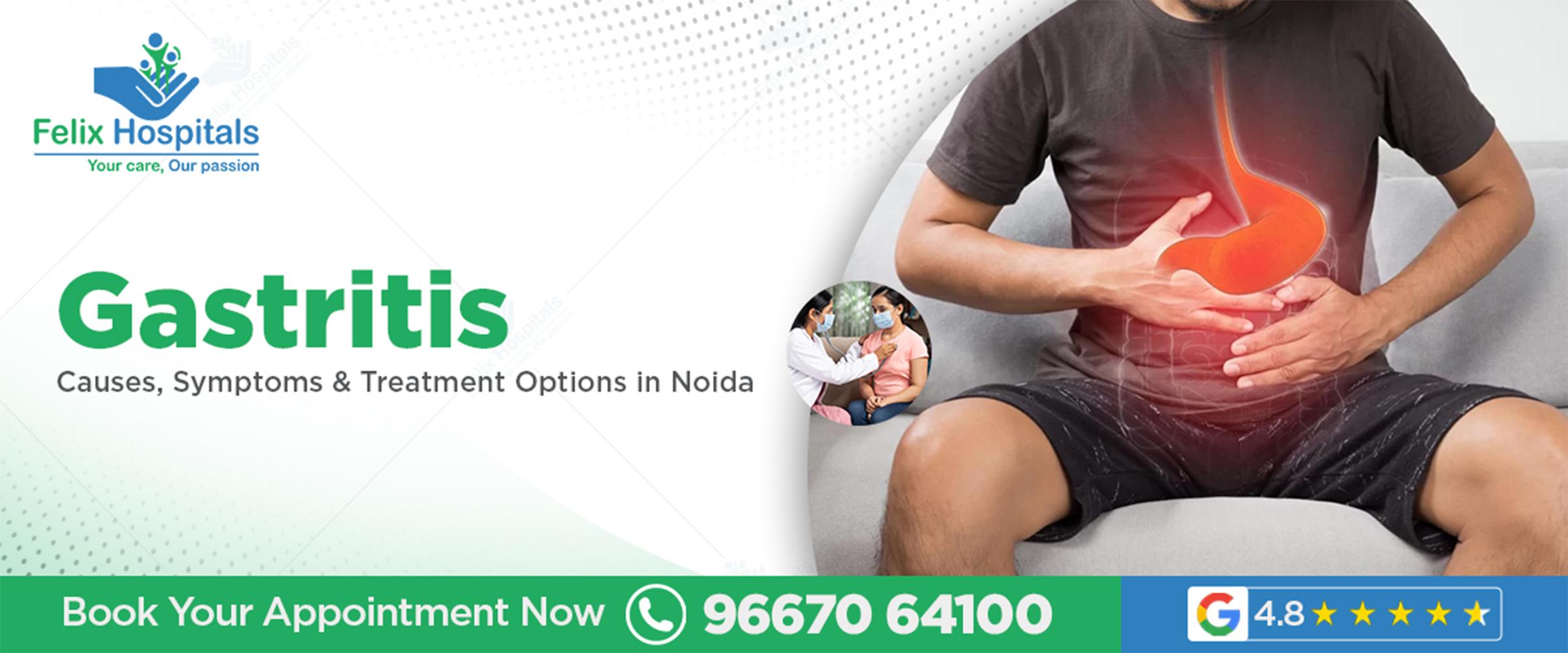
Gastritis refers to inflammation of the stomach lining, which can be acute (sudden) or chronic (long-term). When the stomach lining is irritated or damaged, it affects digestive function and may lead to pain, nausea, and digestive disturbances. Understanding gastritis causes and symptoms helps patients seek timely medical care before complications like ulcers or gastrointestinal bleeding occur.
Several factors contribute to gastritis:
A bacterial infection that weakens the stomach lining and increases acid production, causing irritation and inflammation.
Painkillers such as ibuprofen or aspirin can erode the stomach lining when taken frequently.
Both alcohol and tobacco irritate the stomach lining, promoting inflammation.
Chronic stress, irregular meals, and poor diet contribute to gastritis development.
The immune system mistakenly attacks stomach cells, leading to chronic inflammation.
Prolonged vomiting, bile reflux, and certain medications may also trigger gastritis.
Symptoms vary from mild discomfort to severe pain. Knowing the early signs is important for timely intervention:
Upper abdominal pain or burning sensation
Nausea and vomiting
Loss of appetite
Bloating and indigestion
Belching and heartburn
Black or tarry stools (in severe cases)
Patients experiencing any of these signs should consult a Gastritis specialist doctor in Noida promptly for evaluation. Recognizing acute gastritis symptoms early ensures faster relief and prevents chronic complications.
Accurate gastritis diagnosis involves:
Medical History and Physical Examination – Doctors evaluate symptoms, lifestyle factors, and risk conditions.
Endoscopy – A camera is used to visually examine the stomach lining and identify inflammation or ulcers.
H. pylori Testing – Blood, stool, or breath tests detect the presence of Helicobacter pylori.
Imaging Tests – X-rays with barium can reveal stomach lining abnormalities in some cases.
Lab Tests – Blood tests can detect anemia caused by chronic bleeding from gastritis.
Early chronic gastritis treatment relies on proper diagnosis and identification of the underlying cause.
Treatment varies depending on the type and cause of gastritis.
Antacids: Neutralize stomach acid for immediate relief
Proton Pump Inhibitors (PPIs): Reduce acid production to allow healing
H2 Blockers: Decrease stomach acid and reduce irritation
Antibiotics: Prescribed for H. pylori infections
Avoid spicy, oily, and acidic foods
Eat small, frequent meals to reduce stomach burden
Limit alcohol and quit smoking
Manage stress through meditation or relaxation exercises
Over-the-counter medications may relieve temporary discomfort, but prolonged use without consultation can worsen gastritis. Always seek guidance from a gastritis specialist doctor in Noida.
Chronic gastritis requires continuous monitoring and follow-up. In addition to medications, regular checkups help prevent complications like peptic ulcers or stomach cancer.
Severe cases may require hospitalization for intravenous medications, fluids, and specialized care. A well-equipped Gastritis treatment hospital in Noida provides advanced diagnostic and therapeutic services.
Diet plays a major role in controlling gastritis symptoms:
Include easily digestible foods like rice, oats, and boiled vegetables
Consume lean proteins such as fish, chicken, and legumes
Avoid carbonated drinks, coffee, and acidic juices
Stay hydrated with water and herbal teas
Introduce probiotics to improve gut health
Gastritis diet and lifestyle changes complement medical treatment and enhance recovery.
Preventive measures reduce the risk of gastritis flare-ups:
Avoid excessive alcohol and tobacco use
Limit NSAID consumption unless prescribed
Maintain regular meal timings
Manage stress through yoga, meditation, or exercise
Undergo routine checkups for digestive health
Early attention to warning signs allows timely Gastritis treatment in Noida and reduces long-term complications.
Take medications as prescribed without skipping doses
Follow dietary and lifestyle recommendations strictly
Monitor symptoms and consult a Gastritis specialist doctor in Noida if pain or discomfort persists
Avoid self-medicating for prolonged periods
Attend follow-up appointments to ensure full recovery
With proper care, patients can manage gastritis effectively and maintain a healthy, active life.
Gastritis is a common but manageable condition when identified early. Understanding Gastritis causes and symptoms, seeking timely medical attention, and adhering to prescribed treatment ensures effective management. Whether it is chronic gastritis treatment or dietary modifications, professional guidance is crucial.
Patients seeking advanced care can rely on a Gastritis hospital in Noida for diagnosis, treatment, and long-term management. For consultation with top gastroenterologists and expert care, schedule your visit for Gastritis treatment in Noida today.
Call +91 9667064100 to book an appointment with the best specialists in the region.
Q. 1. What are the common symptoms of gastritis?
Ans. Upper abdominal pain, nausea, bloating, heartburn, and loss of appetite are common signs.
Q. 2. How is gastritis diagnosed?
Ans. Through endoscopy, H. pylori tests, imaging, and lab blood tests.
Q. 3. Can diet alone treat gastritis?
Ans. Diet helps manage symptoms but must be combined with medication for effective treatment.
Q. 4. Is chronic gastritis dangerous?
Ans. If untreated, it can lead to ulcers, bleeding, or increased risk of stomach cancer.
Q. 5. When should I see a gastroenterologist?
Ans. Persistent stomach pain, frequent nausea, or vomiting warrants consultation with a Gastritis specialist doctor in Noida.

Expert gastroenterology care for better digestive health, offering advanced diagnosis and treatment for GERD, IBS, ulcers, and liver disorders.

Experiencing persistent stomach pain, bloating, or acid reflux? These symptoms may require a gastroenterologist’s attention. Early diagnosis is key to treatment.
Understand the factors that influence gastric balloon surgery cost in Noida, including hospital facilities, doctor expertise, and post-procedure care.

हमारी आंखें हमारे जीवन का सबसे महत्वपूर्ण हिस्सा हैं। यह हमारे चारों ओर की दुनिया को देखने, रंगों को पहचानने और दिन-प्रतिदिन की गतिविधियों को करने में मदद करती हैं। लेकिन बदलती जीवनशैली, लंबे समय तक स्क्रीन का उपयोग, अनियमित नींद, तनाव, और विभिन्न स्वास्थ्य समस्याओं के कारण आंखों की बीमारियों का खतरा तेजी से बढ़ रहा है। इनमें से एक गंभीर और अक्सर अनदेखा किया जाने वाला रोग है ग्लूकोमा।
ग्लूकोमा धीरे-धीरे दृष्टि को प्रभावित करता है और कई बार शुरुआती लक्षण दिखाई नहीं देते। अगर समय पर निदान और इलाज न किया जाए, तो यह स्थायी अंधेपन का कारण बन सकता है। इस रोग के बारे में जागरूक होना इसलिए बेहद जरूरी है। नोएडा में ग्लूकोमा डॉक्टर (Glaucoma Doctor in Noida) और आधुनिक तकनीक से लैस आंखों की जांच अस्पताल इस बीमारी का सही निदान और प्रभावी इलाज प्रदान करते हैं।
अधिक जानकारी और आंखों की जांच के लिए कॉल करें: +91 9667064100 और अपनी दृष्टि को सुरक्षित रखें।
ग्लूकोमा एक ऐसी आंखों की बीमारी है जिसमें आंखों के भीतर का दबाव (Intraocular Pressure) बढ़ जाता है। आंख के अंदर हमेशा तरल पदार्थ (Aqueous Humor) बनते और निकलते रहते हैं। यदि यह तरल सही तरीके से बाहर नहीं निकलता, तो आंखों का दबाव बढ़ जाता है।
इस बढ़े हुए दबाव से ऑप्टिक नर्व (Optic Nerve) पर असर पड़ता है, जो दृष्टि को नियंत्रित करती है। धीरे-धीरे ऑप्टिक नर्व को नुकसान पहुँचता है और दृश्य क्षेत्र सीमित होने लगता है। अगर समय पर इलाज न हो, तो यह स्थायी अंधापन का कारण बन सकता है।
ओपन-एंगल ग्लूकोमा (Open-Angle Glaucoma) – यह सबसे सामान्य प्रकार है, जिसमें आंखों का दबाव धीरे-धीरे बढ़ता है और शुरुआती लक्षण दिखाई नहीं देते।
क्लोज़्ड-एंगल ग्लूकोमा (Closed-Angle Glaucoma) – इसमें अचानक गंभीर लक्षण दिखाई देते हैं, जैसे तेज दर्द, धुंधली दृष्टि और आंखों में लालिमा।
ग्लूकोमा शुरूआत में अक्सर साइलेंट ब्लाइंडनेस की तरह होता है। मरीज को शुरू में कोई परेशानी महसूस नहीं होती, लेकिन धीरे-धीरे दृष्टि प्रभावित होने लगती है। इसके लक्षण हैं:
आंखों में धुंधलापन या अस्पष्ट दृष्टि
तेज सिरदर्द या आंखों में दबाव का एहसास
रात में दृष्टि का कम होना
रंगों को पहचानने में कठिनाई
अचानक आंखों में लालिमा या दर्द (क्लोज़्ड-एंगल में)
किसी वस्तु के चारों तरफ हल्की चमक या आभा दिखाई देना
यदि आप उपरोक्त लक्षण महसूस करते हैं, तो ग्लूकोमा डॉक्टर नोएडा से तुरंत संपर्क करना जरूरी है।
ग्लूकोमा के मुख्य कारण आंखों में तरल पदार्थ का असंतुलित प्रवाह है। आंखों के भीतर हमेशा थोड़ी मात्रा में तरल पदार्थ (Aqueous Humor) बना और निकलता रहता है। यदि तरल का बहाव बाधित हो जाता है, तो आंखों का दबाव बढ़ता है और ऑप्टिक नर्व पर दबाव पड़ता है।
उम्र बढ़ना: 40 साल के बाद ग्लूकोमा का खतरा बढ़ जाता है।
परिवार में इतिहास: अगर परिवार में किसी को ग्लूकोमा है, तो जोखिम अधिक होता है।
डायबिटीज और उच्च रक्तचाप: ये आंखों की रक्त वाहिकाओं को प्रभावित करते हैं।
आंख की चोट या सर्जरी: किसी भी चोट से आंखों का दबाव असमान हो सकता है।
दीर्घकालिक दवा का उपयोग: कुछ दवाओं के लंबे समय तक उपयोग से आंखों का दबाव बढ़ सकता है।
ओपन-एंगल ग्लूकोमा: धीरे-धीरे बढ़ता है, शुरुआती लक्षण प्रायः दिखाई नहीं देते।
क्लोज़्ड-एंगल ग्लूकोमा: अचानक गंभीर लक्षण दिखाता है, तेज दर्द और धुंधली दृष्टि।
जन्मजात ग्लूकोमा: बच्चों में दुर्लभ लेकिन गंभीर स्थिति।
सेकंडरी ग्लूकोमा: किसी अन्य बीमारी या आंख की चोट के कारण (Causes of eye injury) उत्पन्न।
ग्लूकोमा का इलाज इसके प्रकार और गंभीरता पर निर्भर करता है।
आंखों की ड्रॉप्स का उपयोग दबाव को नियंत्रित करने के लिए किया जाता है।
कुछ दवाएं तरल के उत्पादन को कम करती हैं, जबकि अन्य बहाव को बढ़ाती हैं।
आंख के तरल बहाव की प्रणाली को सुधारा जाता है।
ओपन या क्लोज़्ड-एंगल ग्लूकोमा में प्रभावी।
गंभीर मामलों में सर्जरी की आवश्यकता होती है।
ट्रैबेकुलेक्टॉमी या अन्य मॉडर्न सर्जिकल तकनीकें आंखों में दबाव कम करती हैं।
नोएडा में आंखों की जांच अस्पताल और अनुभवी ग्लूकोमा डॉक्टर समय पर निदान और प्रभावी उपचार प्रदान करते हैं।
नियमित आंखों की जांच कराएं, विशेषकर 40 साल के बाद।
संतुलित आहार लें, जिसमें विटामिन A और ओमेगा-3 फैटी एसिड शामिल हों।
लंबे समय तक स्क्रीन पर न रहें और हर 2 घंटे में आंखों को आराम दें।
ब्लड प्रेशर और शुगर लेवल नियमित चेक करवाएं।
धूम्रपान और शराब से बचें।
परिवार में यदि किसी को ग्लूकोमा है, तो नियमित फॉलो-अप आवश्यक है।
ग्लूकोमा केवल एक आंखों की सामान्य समस्या नहीं है, बल्कि यह एक गंभीर दृष्टि-संबंधी रोग है जो समय रहते पहचान और उपचार न होने पर स्थायी अंधापन का कारण बन सकता है। इसकी सबसे बड़ी चुनौती यह है कि प्रारंभिक चरणों में इसके कोई स्पष्ट लक्षण दिखाई नहीं देते, जिससे कई लोग इसे नजरअंदाज कर देते हैं। आंखों में हल्का धुंधलापन, रात में दृष्टि में कमी, हल्का सिरदर्द या रंगों को पहचानने में कठिनाई जैसी छोटी-छोटी समस्याएं भी ग्लूकोमा के शुरुआती संकेत हो सकते हैं।
समय पर निदान और उपचार के द्वारा इस रोग को नियंत्रित किया जा सकता है। दवाओं, लेज़र थैरेपी और आवश्यक सर्जिकल उपायों के माध्यम से आंखों में दबाव को नियंत्रित किया जा सकता है और ऑप्टिक नर्व को नुकसान से बचाया जा सकता है। इसके साथ ही नियमित आंखों की जांच, संतुलित आहार, स्क्रीन टाइम पर नियंत्रण और स्वास्थ्य स्थितियों जैसे ब्लड प्रेशर और शुगर का नियंत्रण ग्लूकोमा की रोकथाम में मदद करता है।
नोएडा में अनुभवी ग्लूकोमा डॉक्टर और आधुनिक आंखों की जांच अस्पताल इस रोग का समय पर निदान और प्रभावी इलाज प्रदान करते हैं। विशेषज्ञों के मार्गदर्शन में इलाज से न केवल दृष्टि सुरक्षित रहती है, बल्कि जीवन की गुणवत्ता भी बनी रहती है। इसलिए आंखों की सेहत को कभी नजरअंदाज न करें और शुरुआती लक्षण दिखते ही डॉक्टर से सलाह लें।
सवाल 1. क्या ग्लूकोमा केवल वृद्ध लोगों में होता है?
जवाब: नहीं, ग्लूकोमा किसी भी उम्र में हो सकता है। हालांकि 40 साल से ऊपर लोगों में इसका खतरा अधिक होता है। जन्मजात ग्लूकोमा और secondary ग्लूकोमा बच्चों और युवा लोगों में भी पाया जा सकता है।
सवाल 2. क्या ग्लूकोमा का इलाज पूरी तरह से हो सकता है?
जवाब: ग्लूकोमा का इलाज रोग की गंभीरता पर निर्भर करता है। शुरुआती स्टेज में दवाओं और लेज़र थैरेपी से आंखों के दबाव को नियंत्रित किया जा सकता है। गंभीर मामलों में सर्जरी की आवश्यकता हो सकती है। नियमित फॉलो-अप से दृष्टि को स्थिर रखा जा सकता है।
सवाल 3. क्या ग्लूकोमा में आंखों का लाल होना सामान्य है?
जवाब: आंखों का हल्का लाल होना कभी-कभी सामान्य हो सकता है, लेकिन अगर यह दबाव, दर्द या धुंधली दृष्टि के साथ हो तो यह क्लोज़्ड-एंगल ग्लूकोमा का संकेत हो सकता है। ऐसी स्थिति में तुरंत ग्लूकोमा डॉक्टर नोएडा से संपर्क करें।
सवाल 4. क्या ग्लूकोमा में दृष्टि अचानक खराब हो सकती है?
जवाब: ओपन-एंगल ग्लूकोमा में दृष्टि धीरे-धीरे प्रभावित होती है, जबकि क्लोज़्ड-एंगल ग्लूकोमा में अचानक दृष्टि में धुंधलापन, दर्द और लालिमा देखने को मिल सकती है। ऐसे मामलों में तुरंत आपातकालीन इलाज की आवश्यकता होती है।
सवाल 5. क्या ग्लूकोमा केवल एक आंख को प्रभावित करता है या दोनों?
जवाब: अधिकतर मामलों में दोनों आंखों को प्रभावित कर सकता है, लेकिन शुरुआत में एक आंख अधिक प्रभावित हो सकती है। समय पर इलाज से दोनों आंखों की रोशनी सुरक्षित रखी जा सकती है।
ग्लूकोमा एक गंभीर आंखों की बीमारी है जिसमें ऑप्टिक नर्व को नुकसान पहुंचता है। ऑप्टिक नर्व वह नर्व होती है जो आंख से मस्तिष्क तक दृष्टि की जानकारी पहुंचाती है। ग्लूकोमा का मुख्य कारणं आंखों के अंदर बढ़ा हुआ दबाव (इंट्राओकुलर प्रेशर) है।
ग्लूकोमा में आंख का प्रेशर बढ़कर नजर कमजोर होती है। समय पर इलाज से दृष्टि बचाई जा सकती है।

Subscribe to our
© Copyright 2026. All Rights Reserved by Felix Healthcare Private Limited
Design and development by :


 +(91)9667064100
 Emergency : +(91)9667064100