 WhatsApp
 WhatsApp


Subscribe to our

Let us understand- what is MCh Blood test. MCH, or Mean Corpuscular Hemoglobin, is a measure indicating the average hemoglobin quantity within each red blood cell, responsible for transporting oxygen from the lungs to the cells through the bloodstream. Deviations in MCH levels, identified through blood testing, may signify various issues, ranging from nutrient deficiencies to chronic diseases.
Conducted as part of a Comprehensive Blood Count (CBC), the MCH blood test assesses blood composition, including hematocrit, white blood cells (WBC), platelets, hemoglobin, and red blood cells (RBC). This test offers an overview of overall health. MCH is derived from the hemoglobin value (Hgb) and the RBC count, calculated by dividing Hgb by RCB. The normal MCH range is between 26 and 33 picograms (pg) of hemoglobin per RBC.
Your doctor may discuss MCH levels and what is MCh blood Test when explaining certain blood test results. MCH stands for "mean corpuscular hemoglobin," indicating the average amount of hemoglobin, a protein that transports oxygen, in each red blood cell.
MCH (Mean Corpuscular Hemoglobin) levels gauge the average amount of hemoglobin in each red blood cell, while MCHC blood test (Mean Corpuscular Hemoglobin Concentration) levels measure the average weight of hemoglobin relative to the volume of red blood cells. Both serve as indicators of the health of hemoglobin in the bloodstream. Hemoglobin, a crucial blood protein, facilitates the transport of oxygen to the body's cells and tissues through red blood cells. MCH measures the quantity of hemoglobin in a red blood cell, while the MCHC blood test value signifies the amount of hemoglobin per unit volume.
This blog will delve into the meaning of MCHC in blood tests, elucidate the causes of high MCHC, and unravel the implications of low MCHC levels.
Physicians frequently request a complete blood count (CBC) test to determine an individual's MCH levels. In adults, typically MCH blood test normal range varies from approximately 27 to 31 picograms (pg) per cell, although variations can occur depending on the testing machine and 32 to 36 grams per deciliter (g/dL), or 320 to 360 grams per liter (g/L) for MCHC blood test.
For young children, the reference values differ. Low MCH is indicated by concentrations at or below 27 pg per cell, while high MCH levels are defined by concentrations at 34 pg per cell or higher.
In summary, the answer to what is MCh blood test is that the MCH blood test is a fundamental tool gauging hemoglobin levels in red blood cells, vital for diagnosing and monitoring specific anemias and blood disorders. Typically conducted as part of a complete blood count (CBC), this test aids in identifying underlying health concerns. While comprehending MCH results provides valuable insights into overall health, it's essential to acknowledge that additional testing may be required for a comprehensive understanding of blood health. Overall, the MCH blood test and MCH blood test normal range remains a valuable asset for preserving and enhancing overall health and well-being.
The MCH blood test involves a straightforward process where a small blood sample is taken from a vein in your arm. This sample is usually collected with a needle connected to a syringe or a vacuum tube. The blood is then sent to a laboratory for analysis, where the hemoglobin levels in each red blood cell are measured. Results are typically available within a few days, and your healthcare provider will interpret the results, explaining whether further testing or treatment is necessary.
Additionally, there are now mobile phlebotomy services available for blood draws in the comfort of your home or office. This option is convenient for those who find it challenging to visit a healthcare provider's office or prefer the ease of having blood drawn at home. Services like Speedy Sticks offer mobile blood draw services, and Mobile Labs provide on-site laboratory testing and diagnostics.
A comprehensive blood count test, commonly known as CBC test, aims to provide physicians with a broad understanding of an individual's overall health. This test is valuable for screening multiple health aspects simultaneously, aiding in the diagnosis of conditions like bleeding disorders, infections, and anemia.
CBC tests analyze the three main types of blood cells, offering counts for white cells, red cells, and platelets. A CBC assesses the diverse cellular components constituting your blood, encompassing:
You might undergo a CBC during your annual check-up or as a diagnostic measure for a specific ailment. If you exhibit symptoms indicative of a condition impacting your blood cell count, your doctor may recommend this test.
For the CBC procedure, a nurse inserts a needle into a vein in your arm. The needle is connected to a test tube, collecting the blood, which is later analyzed in a laboratory.
The mean corpuscular hemoglobin (MCH) test is a component of a complete blood count (CBC), a standard blood test used for diagnosing and monitoring various health conditions. A CBC may be conducted during routine health check-ups or when assessing specific health concerns.
Anemia, a prevalent health issue with diverse causes, often prompts a CBC, including an evaluation of MCH. If you exhibit signs or symptoms of anemia, your doctor may order a CBC to confirm the diagnosis and identify the underlying cause.
Causes of Decreased MCH Levels
Several potential factors can contribute to low MCH levels, including:
Abnormal MCH results in blood tests can be a symptom of anemia. Frequently, an insufficient supply of iron leads to anemia, resulting in a low MCH. Iron is essential for the production of hemoglobin in your body.
Pregnancy, blood loss, and weight loss surgery can contribute to a decrease in iron levels, resulting in iron-deficiency anemia or low levels of hemoglobin and MCH..
If you experience iron deficiency anemia, you may notice symptoms such as:
Elevated MCH levels often indicate the presence of macrocytic anemia, where blood cells are larger than normal. This condition can arise due to insufficient levels of vitamin B12 or folic acid in the body.
Additionally, high MCH scores may be linked to the following factors:
Individuals facing increased MCH levels due to macrocytic anemia may exhibit symptoms that follow a specific pattern. While symptoms may not be immediately noticeable, they can gradually intensify over time. Signs of elevated MCH include:
Moreover, those with macrocytic anemia might encounter digestive issues, including loss of appetite, weight loss, and regular diarrhea. It is advisable for individuals experiencing any of these symptoms to consult their doctor promptly.
The approach to addressing unbalanced MCH levels varies for each case.
Dietary Modifications
Doctors may suggest incorporating more iron and vitamin B6 into the diet. Boosting MCH levels can be facilitated by consuming vitamin C and fiber, along with foods rich in iron.
Iron-rich foods include:
For individuals dealing with high MCH levels, incorporating more vitamin B12 and folic acid into the diet is beneficial. While obtaining these nutrients from a diverse and balanced diet is ideal, supplements may also be considered to maintain appropriate levels.
Supplements, such as those containing iron, vitamin B12, vitamin C, and folic acid, can aid in increasing MCH levels when lacking in the diet. However, individuals with MCH imbalances should always consult their doctors before introducing supplements or making significant dietary changes.
Treatment for either low or high MCH levels depends on the root cause. Here's a summary:
Low MCH:
High MCH:
For the best 24-Hour Blood Bank Facility, reach out to Felix Hospitals – Your Lifesaver, Anytime!"
Iron-Enriched Diet
Vitamin B6 Boost
Folate-Enriched Choices
Vitamin B12 Inclusion
Hydration Maintenance
Copper-Rich Selections
Regular Exercise Routine
Incorporating these natural strategies into your lifestyle can significantly boost MCH levels, promoting overall health and enhancing vitality. Remember, investing in your well-being today ensures a healthier and happier life tomorrow.
To your sheer delight, Felix Hospital provides you with the most reasonable and budget-friendly blood test.
In conclusion, here is the answer for what is MCh blood test. The MCH blood test normal range is determined by dividing the total hemoglobin amount by the number of red blood cells in the blood sample. This calculation offers insights into the average hemoglobin content in each red blood cell. The MCH blood test (MCH blood test normal range ) is valuable for diagnosing and monitoring various types of anemia and other blood disorders. Metropolis Labs, a renowned diagnostic and pathology service provider in India, offers a range of tests, including the MCH blood test, to evaluate different facets of an individual's health.
If you are looking for best hospital in Noida, Visit Felix Hospital or Call +(91)9667064100.

Angiography is an important diagnostic test used to detect blockages or narrowing in blood vessels, especially in heart patients.

इस लेख में, हम लिम्फोसाइट्स (lymphocytes in Hindi meaning) का अर्थ, उनके उच्च और निम्न स्तर, कारण, लक्षण, जांच, और उपचार से संबंधित जानकारी प्राप्त करेंगे।
लोग अक्सर कमजोर इम्यूनिटी के कारण बीमार पड़ जाते हैं। ये श्वेत रक्त कोशिकाएं हमें बीमारियों से लड़ने और स्वस्थ होने में मदद करती हैं। इन्हीं श्वेत रक्त कोशिकाओं का एक प्रकार होता है, जिसे लिम्फोसाइट्स (lymphocyte Hindi meaning) कहा जाता है।
इसलिए, इन कोशिकाओं की सही मात्रा में होना अत्यंत महत्वपूर्ण है। डॉक्टर द्वारा नियमित रक्त परीक्षण के दौरान लिम्फोसाइट्स का परीक्षण किया जा सकता है। इन कोशिकाओं का स्तर हर स्थिति में थोड़ा भिन्न हो सकता है।
लिम्फोसाइटोसिस का सही इलाज पाएं — आज ही अनुभवी डॉक्टर से संपर्क करें।
लिम्फोसाइट्स वे व्हाइट ब्लड सेल्स हैं जो हमारे शरीर में मिलते हैं और संक्रमण और बीमारियों से बचाव में महत्वपूर्ण भूमिका निभाते हैं। इन्हें हमारे इम्यून सिस्टम के सैनिक कहा जाता है, जो निरंतर हानिकारक आक्रमणकारियों जैसे कि बैक्टीरिया, वायरस, और अन्य पैथोजन्स को पहचाने में लगे रहते हैं।
लिम्फोसाइट्स (lymphocytes in hindi meaning) अलग-अलग भागों में बनते हैं, जैसे कि बोन मैरो और थाइमस ग्रंथि, और ये पूरे रक्त परिसंचरण और लिम्फेटिक सिस्टम में बटते हैं। जब इन्हें किसी बाहरी पदार्थ से नाकाबूंद करना पड़ता है, तो ये मिलकर उसे पहचानने और समाप्त करने में मदद करते हैं, जिससे हमारा शरीर का रक्षा प्रणाली मजबूत होता है।
एक्सपर्ट सुझाव के लिए कॉल करें +91 9667064100
लिम्फोसाइट्स वे कोशिकाएं हैं जो हमारे शरीर में होती हैं और हमें बीमारियों से बचाने में मदद करती हैं। इन्हें दो प्रकारों में बाँटा गया है - टी लिम्फोसाइट्स (lymphocyte hindi meaning) और बी लिम्फोसाइट्स।
1. टी लिम्फोसाइट्स: ये कोशिकाएं हमारे शरीर में बीमारियों के खिलाफ लड़ने और उन्हें मिटाने में मदद करती हैं। ये शरीर में बनती हैं और जब कोई बीमारी हमारे शरीर में प्रवेश करती है, तो ये उसे पहचानकर मिटाने में मदद करती हैं। इनका जीवनकाल लम्बा होता है।
2. बी लिम्फोसाइट्स: ये भी हमारे शरीर को बीमारियों से बचाने में मदद करती हैं। ये शरीर में बनती हैं और जब कोई बीमारी हमारे शरीर में प्रवेश करती है, तो ये उसे मिटाने में मदद करती हैं। इन्हें हमारे शरीर में रहने वाली दुष्प्रभावित कोशिकाएं पहचानने की क्षमता होती है।
ये सेल्स हमारे बॉडी में इम्यूनिटी (सुरक्षा) बनाने में मदद करते हैं। इनका मुख्य काम होता है कि जब भी कोई बीमारी वाला वायरस या बैक्टीरिया आता है, तो ये उसको पहचान कर उसका मुकाबला करते हैं। इनकी एक महत्वपूर्ण बात ये है कि वे अपने पास्ट में हुई लड़ाइयों को याद रखकर भविष्य में भी त्वरित और प्रभावी तरीके से जवाब दे सकते हैं।
1. बीमारियों को पहचानना: ये सेल्स किसी भी हानिकारक वायरस या बैक्टीरिया को पहचानने में मदद करते हैं।
2. इम्यूनिटी बनाए रखना: इनका काम है हमें पहले से ही सिखा हुआ वायरस या बैक्टीरिया को याद रखकर भविष्य में उनके हमले के लिए तैयार रहना।
3. एंटीबॉडीज बनाए: कुछ लिम्फोसाइट्स, जिन्हें हम बी-सेल्स भी कहते हैं, वे एंटीबॉडीज बनाते हैं। ये एंटीबॉडीज हमारे बॉडी को हमले करने वाले किसी भी हानिकारक तत्व से बचाते हैं।
4. सीधा हमला: कुछ और लिम्फोसाइट्स, जो टी-सेल्स कहलाते हैं, सीधे संक्रमित सेल्स पर हमला करते हैं, जिससे वो और आगे नहीं बढ़ सकते।
लिम्फोसाइट्स गिनती वह प्रक्रिया है जिसमें हम रक्त परीक्षण के बाद अपनी गिनती की सीमा को जानते हैं। इसके लिए हमें डॉक्टर से संपर्क करना चाहिए, ताकि हमें अपने परिणामों का सही समझ मिल सके। क्योंकि यह प्रयोगशाला विभिन्न तरीकों से कोशिकाओं की गिनती करती है, इसलिए परिणाम भिन्न हो सकते हैं।
लिम्फोसाइट (lymphocytes in Hindi meaning) गिनती का मतलब है कि हमारे रक्त में कितने लिम्फोसाइट्स हैं। यह गिनती वयस्कों के लिए और बच्चों के लिए भी अलग-अलग होती है। वयस्कों के लिए सामान्यतः रक्त के प्रति माइक्रोलीटर 1000 से 4800 लिम्फोसाइट्स तक की सीमा होती है, जबकि बच्चों में यह 3000 से 9500 प्रति माइक्रोलीटर रक्त के बीच होता है।
लिम्फोसाइट्स वे शरीर के कारगर होते हैं जो कैंसर, प्रतिरक्षा और ऑटोइम्यूनिटी में महत्वपूर्ण भूमिका निभाते हैं। इन शरीररक्षकों की संख्या को तीन कारकों - कोर्टिसोल, एस्ट्रोजन, और टीएसएच के साथ जोड़ा जा सकता है। यहां लिम्फोसाइट्स की संख्या का सामान्य मापन है:
1. सामान्य लिम्फोसाइट्स स्तर
व्यक्ति की आयु, वंश, लिंग, ऊँचाई, और जीवनशैली पर निर्भर करता है कि लिम्फोसाइट्स की सामान्य संख्या कितनी होनी चाहिए। उदाहरण के लिए:
2. उच्च लिम्फोसाइट्स स्तर
वयस्कों में, अगर प्रति 1 माइक्रोलीटर रक्त में लिम्फोसाइट्स (lymphocyte hindi meaning) 4,800 से अधिक हैं, तो इसे उच्च लिम्फोसाइट्स स्तर माना जाता है। इसे लिम्फोसाइटोसिस भी कहा जाता है, जो आमतौर पर किसी संक्रमण या अंदरूनी बीमारी का संकेत हो सकता है। यह उच्च स्तर शरीर को बीमारियों से बचाने का प्रयास हो सकता है।
3. निम्न लिम्फोसाइट्स स्तर
वयस्कों में, अगर प्रति 1 माइक्रोलीटर रक्त में लिम्फोसाइट्स 1,000 से कम हैं, तो इसे निम्न लिम्फोसाइट्स स्तर माना जाता है।
जब लिम्फोसाइट की संख्या बढ़ जाती है, तो आपको इन लक्षणों का सामना हो सकता है। जैसे:
1. लसीका ग्रंथि में सूजन: आपकी लसीका ग्रंथि में सूजन हो सकती है।
2. रात में ज्यादा पसीना: आपको रात में ज्यादा पसीना आ सकता है।
3. बुखार (Fever): बुखार हो सकता है।
4. पेट में दर्द (Stomach ache) : आपके पेट में दर्द हो सकता है।
5. भूख की कमी: खाने के लिए भूख नहीं लगती है।
6. सांस लेने में दिक्कत: कभी-कभी आपको सांस लेने में भी दिक्कत हो सकती है।
रक्त में लिम्फोसाइट(meaning of lymphocytes in hindi) की कमी को लिम्फोपीनिया या लिम्फोसाइटोपेनिया भी कहा जाता है। यह स्थिति जन्मजात भी हो सकती है या समय के साथ विकसित हो सकती है। इसके लक्षण नीचे दिए गए हैं:
1. बुखार, बहती नाक और खांसी जैसे लक्षण: संक्रमण के लक्षण हो सकते हैं जैसे बुखार, बहती नाक, और खांसी।
2. त्वचा पर दाने या जलन: त्वचा पर दाने हो सकते हैं या जलन हो सकती है।
3. जोड़ों में सूजन और दर्द: जोड़ों में सूजन और दर्द रह सकता है।
4. लसीका पर्व में सूजन: लसीका पर्व में सूजन हो सकती है।
5. काफी लोगों को मुंह में छाले होते हैं: प्लीहा में भी सूजन आ सकती है।
6. पीलिया (Jaundice): यह एक प्रकार का रक्त की कमी है जिससे त्वचा और आँखों का पीलापन हो सकता है।
लिम्फोसाइट स्तर बढ़ने से, टीएसएच और थायरॉयड की स्थिति में बदलाव हो सकता है। लिम्फोसाइट्स (meaning of lymphocytes in hindi) ज्यादा होने के कारण हो सकता है:
1. संक्रमण: यह बहुत आम है, खासकर उन लोगों में जिन्हें हाल ही में संक्रमण हुआ हो, विशेषकर विषाणु संक्रमण।
2. एपस्टीन-बार विषाणु: यह मोनोन्यूक्लिओसिस बीमारी का कारण हो सकता है।
3. साइटोमेगालोवायरस: जो एक प्रकार का दाद वायरस है।
4. काली खांसी: जो संक्रामक श्वसन है, जिसमें विशिष्ट प्रकार की खांसी होती है।
5. एडेनोवायरस: जो श्वसन प्रणाली से जुड़े संक्रमण के लक्षण पैदा करता है।
6. हेपेटाइटिस: जिससे जिगर में सूजन हो सकती है।
7. चिकनपॉक्स: जो त्वचा पर छाले पैदा कर सकता है।
8. कण्ठमाला: जिसमें पेरोटिड लाल ग्रंथियां सूज सकती हैं।
9. खसरा: एक संक्रमण बीमारी है जो रुवी जीवाणु से हो सकती है।
10. एचआईवी: यह भी एक संक्रमण है जो शरीर की प्रतिरक्षा को कमजोर कर सकता है।
1. दीर्घकालिक लिम्फोसाइटिक ल्यूकेमिया (एक प्रकार का रक्त कैंसर)
2. नॉन-हॉजकिन लिंफोमा (लसीका तंत्र में विकसित होनेवाला एक रक्त कैंसर का समूह)
3. तीव्र लसीका ब्लास्टिक ल्यूकेमिया (कैंसर जो रक्त और अस्थि-मज्जा को प्रभावित करता है)
4. बड़े दानेदार लिम्फोसाइटिक ल्यूकेमिया (दुर्लभ प्रकार का कैंसर जो सफेद रक्त कोशिकाओं को प्रभावित करता है)
इसके अलावा, लिम्फोसाइट्स (meaning of lymphocytes in hindi) के उच्च स्तर से जुड़ी कुछ चिकित्सा स्थितियों में जो लंबे समय तक सूजन बनी रहती है, उनमें भी देखा जा सकता है, जैसे:
1. संधिशोथ
2. किसी नई दवा की प्रतिकूल प्रतिक्रिया
3. आघात जैसी गंभीर चिकित्सा बीमारी
4. तिल्ली हटाने के बाद
5. धूम्रपान
6. किसी चिकित्सा आपात स्थिति से संबंधित तनाव
7. स्व-प्रतिरक्षी रोग
लिम्फोसाइट्स (lymphocytes in hindi meaning) की कमी के कारण, आपकी टीएसएच स्तर जो थायराइड गतिविधि को नियंत्रित करता है, बिगड़ सकता है। लिम्फोसाइट्स कम होने के कई कारण हो सकते हैं। सबसे सामान्य कारण हैं संक्रमण, दवाएं, और पोषण संबंधी कमियां।
संक्रमण - विभिन्न प्रकार के जीवाणु, विषाणु, परजीवी और कवक संक्रमण लिम्फोसाइट्स की कमी का कारण बन सकते हैं। उदाहरण के लिए:
1. एचआईवी और एड्स
2. श्लैष्मिक ज्वर (जिसमें खांसी और बुखार होता है)
3. कोविड-19
4. जिगर में सूजन
5. मलेरिया (Malayria) (मच्छर से होने वाला विषमज्वर)
6. खसरा
7. न्यूमोनिया pneumonia (फेफड़ों की सूजन और संक्रमण)
8. क्षय रोग (Tuberculosis)
कुपोषण - आहार में पोषक तत्वों की कमी, जैसे प्रोभूजिन, विटामिन बी-१२, फोलिक एसिड, आदि, भी लिम्फोसाइट्स की कमी का कारण बन सकती है।
वंशानुगत बीमारियाएं - वह बीमारियां जो किसी के जीवन में उनके माता-पिता से आनुवंशिक रूप से मिलती हैं, वे बहुत नीचे दी गई हैं:
1. गतिभंग रक्त वाहिनी विस्तार (एटैक्सिया-टेलैंगिएक्टेसिया): यह एक बहुत दुर्लभ आनुवंशिक रोग है जो तंत्रिका तंत्र, प्रतिरक्षा प्रणाली, और कई अन्य शरीर के प्रणालियों को प्रभावित कर सकता है।
2. रोगक्षम-अपर्याप्तता: यह एक बीमारी है जिसमें शरीर की प्रतिरक्षा प्रणाली कमजोर होती है, जिससे बीमारियों के खिलाफ सामान्य प्रतिरक्षा कमजोर हो जाती है।
3. डि जॉर्ज सिंड्रोम: यह एक आनुवंशिक रोग है जिसमें क्रोमोसोम २२ का एक हिस्सा अनुपस्थित होता है।
4. गंभीर संयुक्त रोगक्षम-अपर्याप्तता बहुरोग: यह एक बहुरोग है जिसमें बच्चे की पूरी तरह से ठीक प्रकार से बनने में मुश्किलें होती हैं।
5. विस्कॉट-एल्ड्रिच सिंड्रोम: यह एक दुर्लभ आनुवंशिक विकार है जिसमें रक्त के थक्के बनाने की क्षमता कम हो जाती है।
स्व-प्रतिरक्षित रोग - यह होता है जब हमारी प्रतिरक्षा प्रणाली हमारे ही स्वस्थ कोशिकाओं पर हमला करती है, जिसमें लिम्फोसाइट्स भी शामिल होते हैं। उदाहरण के रूप में:
1. ल्यूपस (सिस्तमिक लुपस एरिथेमेटोसस): इसमें त्वचा पर चकत्ते, जोड़ों में दर्द, बुखार, और अंग क्षति होती है।
2. सारकॉइडोसिस: इसमें फेफड़ों, लसीकापर्व, त्वचा, आंखों और शरीर के अन्य हिस्सों में गांठ बनते हैं।
3. मायास्थेनिया ग्रेविस: इससे नर स्नायु प्रणाली के ग्रसित होने से मांसपेशियों में कमजोरी होती है।
4. रियुमेटोइड गठिया: इसमें प्रतिरक्षा प्रणाली हाथ-पैर या पीठ के जोड़ों को अस्तर करने वाले ऊतक को निशाना बनाती है।
दवा और उपचार के दुष्प्रभाव - कुछ रोगों के इलाज में काम आने वाली दवाएं, जैसे कि कैंसर रोग या स्व-प्रतिरक्षित रोग के इलाज के लिए कुछ दवाएं, लिम्फोसाइट्स को कम कर सकती हैं। जैसे:
1. अस्थि-मज्जा प्रत्यारोपण
2. इम्यूनो दमनकारी दवाएं (जैसे कि कॉर्टिकोस्टेरॉइड)
3. विकिरण चिकित्सा
4. रसायन चिकित्सा
लिम्फोसाइट्स (lymphocytes in hindi meaning) के इलाज के लिए, यदि आपके शरीर किसी रोगाणु के साथ लड़ रहा है और श्वेत रक्त कोशिकाएं बना रहा है, तो आपको इसके लिए कोई विशेष उपचार की आवश्यकता नहीं होती है। समय के साथ, प्रतिरक्षा प्रणाली इस स्थिति को सुधारती है। यदि सफेद रक्त कोशिकाओं में बढ़ोतरी किसी गंभीर बीमारी की ओर संकेत कर रही है, तो चिकित्सक आपको उच्च लिम्फोसाइट स्तर के कारणों और उपचार के विकल्पों के बारे में बताएंगे।
उच्च लिम्फोसाइट्स (lymphocytes normal range in hindi) के इलाज में, लिम्फोसाइट्स के उच्च स्तर को नियंत्रित करने के लिए कई उपाय हो सकते हैं:
1. संक्रमणों का समाधान - उच्च लिम्फोसाइट स्तर के कारण होने वाले संक्रमणों के इलाज के लिए विशेष एंटीबायोटिक या एंटीवायरल दवाएं की जा सकती हैं।
2. ऑटोइम्यून स्थितियों का प्रबंधन - ऑटोइम्यून विकारों के लिए दवाओं का प्रबंधन किया जा सकता है, जो लिम्फोसाइट्स की अत्यधिक गिनती को कम कर सकती हैं।
3. तनाव कम करना - योग, ध्यान और व्यायाम जैसी तकनीकें तनाव को कम करके लिम्फोसाइट्स की अत्यधिक गिनती को नियंत्रित करने में मदद कर सकती हैं।
4. दवा समायोजन - कुछ दवाओं के कारण गिनती बढ़ जाने पर, डॉक्टर खुराक को समायोजित कर सकते हैं या विकल्प प्रस्तुत कर सकते हैं।
जब कोई व्यक्ति किसी अन्य बीमारी के निदान के दौरान अचानक अधिक या कम लिम्फोसाइट्स (meaning of lymphocytes in Hindi) की गिनती पाता है, तो उसे डॉक्टर के पास जाना चाहिए। डॉक्टर से परीक्षण के परिणामों की चर्चा करना बहुत महत्वपूर्ण है। उच्च या निम्न लिम्फोसाइट गिनती से रोग के स्रोत का पता लगा जा सकता है।
अक्सर कई हफ्तों के दौरान लगातार परीक्षण से यह पता चलता है कि लिम्फोसाइटोसिस कम हो गया है। यदि लिम्फोसाइटोसिस बनी रहती है, तो विशेष रक्त परीक्षण फायदेमंद हो सकते हैं।
यदि समस्या बिगड़ती है या कारण स्पष्ट नहीं है, तो डॉक्टर हेमेटोलॉजिस्ट, जो रक्त विकारों पर ध्यान केंद्रित करता है, को देखने की सलाह दे सकता है। NCBI के एक लेख में कहा गया है कि लिम्फोसाइटों (meaning of lymphocytes in Hindi) की उच्च संख्या (ALC 30000 कोशिकाओं/माइक्रोलीटर से अधिक) या जो तेजी से बढ़ रही है, उन्हें हेमेटोलॉजिस्ट से परामर्श की आवश्यकता होती है।
यदि आप नोएडा में सर्वश्रेष्ठ अस्पताल की तलाश में हैं, तो फेलिक्स अस्पताल जाएँ या +(91)9667064100 पर कॉल करें।,
For those experiencing such symptoms, seeking care from the best hospital for pulmonology treatment in Noida ensures you receive timely and expert diagnosis and treatment.
Blood cancer affects the production and function of blood cells. Early detection, accurate diagnosis, and advanced treatments significantly improve survival rates and patient outcomes in Noida and beyond.

Are you feeling pain because of a small tear near your back passage, also known as an anal fissure? If so, it's important to find the right treatment. In Noida, there are many options to think about. From making lifestyle changes to having a special kind of surgery, the goal is to make the pain better and help you feel well. Experienced doctors who specialize in this area can help you figure out the best treatment, what might happen, and let you make choices about your health.
Start a journey to feel better and more comfortable. Noida has good hospitals and doctors who are skilled at fixing this problem. Anal Fissure Surgery Cost in Noida is fair, so you can get good care without spending too much. Whether you need a simple treatment or something more involved, Noida has good options that are affordable and can really help you feel better.
Remember, the cost of fissure surgery might be different based on things like the kind of treatment, how complicated your case is, what the hospital is like, the fees for the specialist doctor, and how you're taken care of after the surgery.
Book your consultation with us today with a simple click or call us at +91 9667064100
| Starting Cost | ₹35,000 |
| Average Cost | ₹45,500 |
| Maximum Cost | ₹50,000 |
Looking for affordable anal fissure surgery cost (or Fissure Operation Cost) in Noida. Consult Felix Hospital's top specialists We prioritize exceptional care while ensuring affordability. Our experienced specialists and super specialists doctors and dedicated staff are committed to delivering outstanding treatment that aligns with your budget. Begin your path to relief with confidence, knowing you're receiving top-notch care tailored to your needs.
An anal fissure refers to a small tear in the lining of the anal canal, commonly stemming from constipation, straining during bowel movements, or chronic diarrhea. Individuals often experience intense, sharp pain during bowel movements, accompanied by the passage of bright red blood. If left untreated, fissures can become chronic. These tears are prevalent and can affect both men and women of all ages.
In most cases, surgical intervention is unnecessary for anal fissures. Upon diagnosis, our team collaborates to create a tailored treatment plan. Warm baths can provide relief and enhance blood circulation. Medications, including fiber therapy, aim to modify stool consistency and facilitate fissure healing. Should these measures prove insufficient, sphincter relaxants may be prescribed. Surgical options are reserved for cases where other approaches fail.
While rare, an anal fissure may signal an underlying disorder such as inflammatory bowel disease (IBD), sexually transmitted disease (STD), or cancer. If the diagnosis remains unclear, additional tests like an exam under anesthesia or colonoscopy may be required to rule out other conditions.
Surgical treatment is typically reserved for acute anal fissures that continue to show symptoms after 4-6 weeks of medical therapy and for chronic anal fissures. The different types of anal fissure surgery cost (or Fissure Operation Cost) in Noida are as follows:
For surgical treatment of an anal fissure, administering an enema on the morning of the procedure is adequate bowel preparation. In cases where the fissure causes too much pain, the enema may be omitted.
Botulinum toxin (BOTOX®) can be employed to address both acute and chronic anal fissures. It is injected directly into the internal anal sphincter, essentially performing a chemical sphincterotomy. The effects last approximately 3 months until nerve endings regenerate. This period may allow acute fissures (and sometimes chronic fissures) to heal and symptoms to subside, with success rates around 50%.
Lateral internal sphincterotomy is the current preferred surgical procedure for anal fissures. It can be performed with the patient under general or spinal anesthesia. When dealing with a chronic anal fissure, the surgeon may choose to perform a fissurectomy alongside the lateral sphincterotomy.
In cases where long-standing chronic fissures do not heal despite an adequate sphincterotomy, an advancement flap may be necessary to cover the mucosal defect. This procedure can be conducted either concurrently with the sphincterotomy if the surgeon anticipates the fissure not healing or as a subsequent procedure if healing does not occur.
Pain from the fissure begins to diminish almost immediately. The only postoperative restrictions stem from the anesthesia, and many patients can resume normal activities the following day.
Choosing the right anal fissure treatment cost involves considering severity, medical guidance, and personal choices. Expert consultation will help determine the best approach, while cost of fissure surgery estimates vary by location and facility. For personalized anal fissure surgery cost (or Fissure Operation Cost) details, contact Felix Hospital at +91 9667064100, and start your path to relief and well-being.
Your health insurance policy can alleviate the financial burden of treating an illness like a fissure. Covered fissure treatment cost/cost of fissure surgery includes:
If experiencing pain during bowel movements or noticing blood on stools or toilet paper after bowel movements, seeking prompt medical attention is crucial.
The diagnosis of anal fissures typically involves a combination of medical history, physical examination, and sometimes additional diagnostic procedures. Here's how anal fissures are diagnosed:
Medical History:
Physical Examination:
Digital Rectal Examination (DRE):
Anoscopy:
Colonoscopy
Biopsy
It's essential to consult with a healthcare professional for a proper diagnosis and treatment plan tailored to your specific situation. If you suspect you have an anal fissure or are experiencing symptoms, seek medical attention promptly for an accurate diagnosis and appropriate care.
Medical Evaluation:
Physical Examination:
Diagnostic Tests:
Discussion of Treatment Options:
Fasting:
Medication Adjustment:
Hygiene Practices:
Pain Management:
Dietary Recommendations:
Hygiene Instructions:
Activity Restrictions:
Follow-up Appointments:
Stool Softeners or Fiber Supplements:
It's essential to follow your healthcare provider's instructions carefully and attend all follow-up appointments to ensure a smooth recovery and optimal healing. Individual preparations may vary based on the specific surgical procedure and the patient's overall health. Always consult with your healthcare provider for personalized advice.
Pain Management:
Hygiene Practices:
Dietary Adjustments:
Stool Softeners or Fiber Supplements:
Activity Restrictions:
Follow-up Appointments:
Return to Work and Normal Activities:
Resuming Sexual Activity:
Monitoring for Complications:
Long-Term Preventive Measures:
Psychological Support:
Understanding these risk factors aids in identifying individuals who may be more susceptible to anal fistulas and enables proactive measures for prevention and management. Need to know the precise cost for anal fisure surgery?
Choosing Felix Hospital for Anal Fissure Treatment means opting for a comprehensive healthcare experience. Our features include skilled surgeons, advanced facilities, a holistic approach to care, a range of multi and supers specialty services, accreditation for safety and quality, a highly competent medical team, positive patient feedback, a convenient location, cost-effective fissure treatment, complimentary pick and drop services, and state-of-the-art amenities. We also accept both private and government health insurance.
Felix Hospital, we transform the management of anal fissures into a manageable condition through contemporary treatments. Despite the potential challenges in diagnosis, treatment, and recovery, our diverse treatment options cater to individuals of all ages and varying severity levels. Post-treatment, our healthcare providers offer crucial advice on diet, hygiene, and exercises to ensure prompt healing. Your journey to recovery is our priority.
Book an Online Appointment: https://www.felixhospital.com/contact

Anal fissures, perianal abscesses, and anal fistulas are common anorectal disorders that can significantly impact an individual's quality of life.

फिशर के कारण, लक्षण और उपाय" एक संक्षिप्त मार्गदर्शिका है जो फिशर (गुदा裂) के विषय में जानकारी प्रदान करती है। इसमें फिशर के प्रमुख कारण, जैसे कब्ज, जन्म, या अन्य स्वास्थ्य समस्याएं

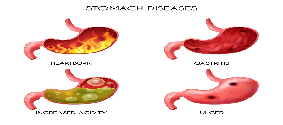
Gastric problem in stomach is a prevalent state that can result in uneasiness and ache. It pertains to any disruption in the normal functioning of the stomach, resulting in different signs and complications. Stomach issues can vary from moderate to intense, impacting people of every age category.
Book Your Appointment One click at +91 9667064100
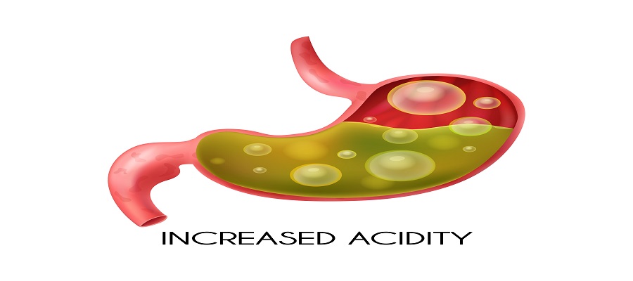
Gastrointestinal issues emerge as a result of multiple factors, including dietary habits, lifestyle preferences, and underlying health conditions. These problems can result in inflammation of the stomach, commonly referred to as gastritis. Gastritis manifests when the protective layer of the stomach becomes irritated or inflamed, giving rise to discomfort and associated indicators. When an individual consumes food or beverages, they also ingest air. To understand what the gastric problem in the stomach is, know that it leads to the creation of gas, which is primarily expelled through burping. Occasionally, certain carbohydrates, fibers, and sugars that are fermented by bacteria do not undergo proper digestion in our digestive system.
While this may appear embarrassing, it is a natural process and there is no need to feel ashamed. Our body performs this function daily in order to maintain the smooth functioning of our internal systems. Were you aware that an individual releases gas approximately 20 times each day? The release of gases primarily occurs before or after meals.
To understand what is gastric problem in stomach, Symptoms of gastric problem may include:
Abdomen discomfort: This is among the frequently experienced indications of gastric issues. The discomfort can vary from a mild throbbing to an intense, burning sensation in the upper abdomen. It may worsen after eating, especially if the stomach is empty.
Heartburn: Famously referred to as acid indigestion, heartburn is a burning sensation in the chest or upper abdomen, often caused by stomach acid flowing back into the esophagus. This prevalent sign of GERD can be incited by specific food items, reclining after eating, or leaning forward.
Nausea and vomiting: To understand what is gastric problem in stomach is, know that it can occasionally trigger sensations of queasiness and might result in vomiting. This may be more prominent in conditions like gastritis or gastroenteritis (stomach flu).
Gas and bloating: Excessive production of gas and bloating are frequently experienced symptoms of gastric problems. This can lead to uneasiness, a sense of being full, and may also be accompanied by burping or releasing gas.
Changes in bowel movements: Stomach problems can impact the regularity of bowel movements, resulting in symptoms like diarrhea or constipation. These changes in stool consistency might be triggered by inflammation or an infection in the gastrointestinal tract.
Loss of appetite: Loss of appetite is one of the main symptoms of gastric problems.
Certain people with stomach problems might encounter a reduced appetite or feel full quickly after eating only a small amount of food. This phenomenon can lead to inadvertent weight reduction as time passes.
Fatigue and weakness: If your gastric problem in the stomach sticks around for a long time, it could mean your body isn't getting all the important nutrients it needs. This can make you feel tired, weak, or uncomfortable. Fatigue and weakness is one of the main symptoms of gastric problems.
There are numerous potential causes of gastric issues or gastric problems in stomach, and it is crucial to acknowledge that individual experiences may vary. Here's a basic look at some common things that can lead to gastric issues and this will help in understanding what is gastric problem in stomach: -
Poor Eating Habits: Consumption of an unhealthy diet is a major factor that contributes to gastric issues. The intake of excessive amounts of fatty, greasy, or spicy foods can cause irritation to the stomach lining, resulting in indigestion, acid reflux, and heartburn. Also, eating too fast or not chewing well can mess up digestion.
Helicobacter Pylori Infection: Helicobacter pylori, also known as H. pylori, is a bacterium that can infect the lining of the stomach and cause stomach inflammation gastritis. Typically, this bacterium is contracted through consuming contaminated food or water, or by having close contact with someone who is infected. An H. pylori infection can result in inflammation, gastritis, peptic ulcers, and an elevated risk of developing gastric cancer.
Medications: Certain medications, such as nonsteroidal anti-inflammatory drugs (NSAIDs) like aspirin or ibuprofen, can irritate the stomach lining and disrupt the protective mucus barrier. Using these medicines excessively or for an extended period of time can increase the risk of experiencing gastrointestinal issues, such as stomach ulcers and bleeding.
Stress and Anxiety: Emotional stress and anxiety can directly affect the digestive system. When a person is stressed, their body releases stress hormones, which can disrupt digestion, increase the production of stomach acid, and delay stomach emptying. Chronic stress can contribute to long-term gastric problems, such as functional dyspepsia or irritable bowel syndrome (IBS).
Smoking and Alcohol Consumption: Both smoking and excessive alcohol intake can irritate the lining of the stomach, resulting in gastritis and an increased likelihood of developing gastric ulcers. Moreover, smoking weakens the lower esophageal sphincter, which enables stomach acid to flow back into the esophagus, causing symptoms of acid reflux.
Genetic Factors: Some people might inherit a tendency for certain stomach problems due to their genes. For instance, specific gene differences can raise the chance of having conditions like GERD or peptic ulcers.
Hormonal Changes: Hormonal changes that occur during pregnancy or menopause can impact the functioning of the digestive system and contribute to gastric issues. Pregnancy hormones can cause the muscles in the digestive tract to relax, which in turn slows down digestion and increases the chances of experiencing acid reflux.
Gastric issues that need quick treatment can be different in how serious they are. It is really important to understand what is gastric problem in stomach, find them early and take care of them fast to prevent any big issues by visiting the best gastroenterologist hospital. Here is some basic information about gastric issues that might need quick treatment:-
Gastric Ulcers: Gastric ulcers are open sores that form on the lining of the stomach. If they are not treated, they can worsen and cause serious issues such as bleeding or holes in the stomach. Immediate treatment for gastric ulcers may include medications that decrease the production of stomach acid (e.g., proton pump inhibitors) and antibiotics if there is an underlying bacterial infection (Helicobacter pylori). In more severe cases, hospitalization may be required for observation and additional interventions.
Stomach Inflammation Gastritis: Stomach inflammation gastritis, typically caused by factors such as excessive alcohol consumption, certain medications, infections, or autoimmune disorders. Although not all cases of gastritis demand immediate treatment, severe acute gastritis or chronic gastritis accompanied by intense symptoms (such as severe abdominal pain, vomiting blood, or black stools) may necessitate urgent attention. Doctors can use medicines to lower stomach acid, antibiotics for infections, and suggest changes in your diet.
Gastroenteritis: Stomach flu, or gastroenteritis, usually happens because of a virus or bacteria. It can result in symptoms such as vomiting, diarrhea, abdominal cramps, and dehydration. Most times, it gets better on its own with rest and drinking enough liquids. There are occasions where immediate treatment may be necessary. Treatment for gastroenteritis can include using oral rehydration solutions or, in more severe scenarios, intravenous fluids administered in a hospital setting.
Gastric Obstruction: Gastric obstruction happens when something blocks the stomach or the initial section of the small intestine (duodenum). This blockage can be from scar tissue, tumors, or certain stomach issues. Signs may include strong stomach pain, feeling really full, not being able to pass gas or poop, and vomiting. A complete gastric obstruction can be a medical emergency that necessitates immediate treatment, potentially involving surgical intervention, to reduce the blockage and restore normal digestive function.
Perforated Peptic Ulcer: A perforated peptic ulcer is a complication of gastric ulcers wherein the ulcer completely penetrates the stomach or duodenal wall. This can result in abrupt and intense abdominal pain that often extends to the back, accompanied by other symptoms of an acute abdomen, such as vomiting, pale appearance, and rapid heartbeat. This needs urgent medical help. Treatment usually means fixing the hole with surgery and taking care of you afterward.
It begins with a burning ache or discomfort (indigestion) in your upper belly and sometimes in the food pipe (esophagus). This feeling might get either worse or better after eating. Symptoms of stomach inflammation gastritis are- nausea, vomiting, a full feeling in the upper belly after eating, heaviness in the stomach, or tiredness can accompany gastric pain.
Your recovery time can vary from 1 week to a maximum of 4 weeks, depending on how serious your situation is. If your gastric problem is mostly from indigestion, you might even feel better in a day with the right medicine.
Eating foods that provide important nutrients like vitamins, minerals, and antioxidants can make a bloated stomach worse and increase gastric pain. It's recommended to avoid foods containing:
Complex sugars
Fructose
Lactose
Insoluble fiber
Starch
Managing stomach problems typically includes making lifestyle adjustments, taking medications, and, in certain instances, considering surgical options. Let's discuss this in detail:-
Lifestyle Adjustments:
Diet:
Eat healthy to control stomach issues.
Avoid or limit spicy, acidic, and fried foods.
Choose a balanced diet with fruits, veggies, whole grains, lean proteins, and low-fat dairy.
Meal Habits:
Have smaller meals more often to lighten the load on your stomach.
Avoid lying down right after eating to prevent acid reflux.
Stress Management:
Manage stress to improve stomach symptoms.
Use techniques like exercise, yoga, meditation, or deep breathing.
Medications:
Antacids:
Use antacids to neutralize stomach acid.
Get temporary relief from heartburn and indigestion.
Proton Pump Inhibitors (PPIs):
Medicines that lower stomach acid production.
Often given for problems like GERD (acid reflux) and stomach ulcers.
H2 Blockers:
Take H2 blockers to decrease stomach acid.
Use them for conditions like gastritis and gastric ulcers.
Antibiotics:
Use antibiotics if a bacterial infection, like H. pylori, causes stomach problems.
Follow a prescribed antibiotic course.
Surgical Interventions:
Endoscopic Procedures:
Use endoscopy to find and treat stomach issues.
It's a simple procedure where a flexible tube with a light and camera checks the stomach and esophagus.
Helpful for tasks like removing polyps, stopping bleeding, or widening strictures.
Surgery to Fix the Issue:
Surgery might be suggested if medicines and lifestyle changes don't work.
For problems like perforated ulcers or bad GERD that doesn't get better with other treatment of gastric problems.
Surgical repair could be needed in these cases.
When you have problems with your stomach or digestion, it's essential to see a gastroenterologist to understand what is gastric problem in stomach and treatment of gastric problems. Here are situations when you should consider consulting a gastroenterologist:
Digestive Problems: If you are experiencing persistent or recurring digestive problems, it is advisable to consult a gastroenterologist. These problems may include, but are not limited to, chronic diarrhea, constipation, abdominal pain, bloating, heartburn, difficulty swallowing, or changes in bowel movement patterns.
Gastrointestinal Bleeding: If you notice blood in your stool or vomit, it could indicate gastrointestinal bleeding. This condition can be caused by various factors like ulcers, hemorrhoids, polyps, inflammatory bowel disease, or even cancer. To diagnose and treat the underlying cause, it is recommended to consult a gastroenterologist.
Abnormal Screening Tests: If you have undergone routine health screenings such as stool tests (for occult blood), colonoscopy, endoscopy, or other diagnostic tests that reveal abnormalities, a gastroenterologist can offer further evaluation and assist you in navigating treatment options, if necessary.
Liver or Pancreatic Diseases: Liver or pancreatic diseases, such as viral hepatitis (hepatitis B and C), cirrhosis, fatty liver, chronic pancreatitis, or pancreatic cancer, often necessitate the knowledge and skills of a gastroenterologist. These medical professionals are adept in effectively diagnosing, managing, and treating such conditions.
Inflammatory Bowel Disease (IBD): Conditions such as Crohn's disease and ulcerative colitis are chronic inflammatory bowel diseases that necessitate specialized care from a gastroenterologist. These conditions can result in severe abdominal pain, diarrhea, rectal bleeding, weight loss, and malnutrition. Seeking assistance from a gastroenterologist can assist in managing the symptoms, preventing complications, and enhancing your overall quality of life.
Gallbladder and Biliary Tract Issues: If you are experiencing problems with your gallbladder or biliary system, such as gallstones, biliary obstructions, or bile duct infections, it is important to consult a gastroenterologist. They can offer an accurate diagnosis and suggest suitable treatments, including surgical interventions if required.
Unexplained weight loss or nutritional deficiencies: If you're losing weight without a clear reason or dealing with nutrient problems even though you eat well, a gastroenterologist can assess your digestive system to identify potential causes, such as malabsorption syndromes or certain gastrointestinal disorders.
Don't eat junk food.
Engage in physical activity.
Say "no" to tobacco and smoking.
Choose healthy foods.
Eat regularly.
In conclusion, taking care of gastric problems is important for overall health. While gastric issues in the stomach are common, if you frequently experience symptoms such as burping, gas, bloating, or stomach pain, it’s advisable to consult a doctor. Sometimes, gas or discomfort may indicate an underlying health concern. Eating healthy foods, maintaining a balanced lifestyle, and avoiding bad habits help manage stomach issues effectively. Seeking advice from the top gastroenterologists in Noida ensures accurate diagnosis and expert treatment. By focusing on digestive health, you can enjoy better digestion and a happier, healthier life.
Understand the factors that influence gastric balloon surgery cost in Noida, including hospital facilities, doctor expertise, and post-procedure care.

Expert gastroenterology care for better digestive health, offering advanced diagnosis and treatment for GERD, IBS, ulcers, and liver disorders.
Understand the factors that influence endoscopy procedure cost in Noida. Learn about the types, preparation, and benefits of choosing the best multi-speciality hospital in Noida for accurate diagnosis.

Appendix surgery, also known as appendectomy, is a common emergency procedure performed to treat appendicitis.

The full form of PCOD and PCOS is Polycystic Ovary Syndrome (PCOS) and Polycystic Ovary Disorder (PCOD). It is widespread hormonal disorders that affect the lives of countless women worldwide. Both conditions have significant impacts on a woman's reproductive health and overall well-being. Polycystic Ovarian Disorder (PCOD) is a medical condition in which a woman's reproductive organs excessively produce immature or partially mature eggs during the reproductive age. These eggs then develop into cysts in the ovaries over time. As a result, the ovaries become enlarged and produce excessive quantities of male hormones (androgen), which can lead to fertility issues, irregular menstrual cycles, undesired weight increase, and various other health complications.
Book Your Appointment One click at +91 9667064100
The full form of PCOD, which stands for Polycystic Ovary Disorder, refers to a complex condition characterized by the presence of multiple cysts on the ovaries. Although these cysts are small, they can disrupt hormonal balance, resulting in a range of symptoms that affect the menstrual cycle, fertility, and overall well-being. Although it is quite common, the majority of women remain unaware of the indications and symptoms of PCOD in females, the consequences of this condition, and the available PCOD Problem treatment alternatives (solution for PCOD problem). Hence, let us delve into it more profoundly.
PCOD can result in various issues and difficulties, impacting not just the reproductive system but also one's overall well-being. To gain a deeper understanding, let us examine the potential problems that PCOD may cause:
Menstrual Irregularities: One of the main symptoms of pcod problem in females is inconsistent menstruation patterns. Individuals with PCOD may encounter occasional or extended periods, or even completely miss periods due to irregular ovulation. Hormonal imbalances disturb the regular menstrual cycle, making it difficult for individuals to predict when they will ovulate or when their periods will happen.
Infertility: PCOD is a primary factor contributing to female infertility. The imbalances in hormones and irregular ovulation associated with PCOD can make it difficult for eggs to mature and be released for fertilization. Additionally, it can lead to the development of numerous ovarian cysts, which further disrupt ovulation. Women with PCOD may encounter difficulties conceiving naturally and might seek medical assistance,i.e. PCOD Problem treatment, such as fertility treatments or therapy to induce ovulation.
Metabolic issues: There is a strong correlation between metabolic difficulties and PCOD. PCOD (Fullform of PCOD- Polycystic Ovary Disorder) is linked to conditions like insulin resistance and obesity. Insulin resistance occurs when cells do not properly respond to insulin, leading to higher insulin levels in the blood. This can contribute to an increase in body weight and create challenges in maintaining proper blood sugar levels, which may ultimately lead to the onset of type 2 diabetes.
Weight Gain: Many women experiencing PCOD encounter difficulties in managing their weight as a consequence of hormonal irregularities and the body's reduced sensitivity to insulin. The high levels of insulin can encourage the heightened production of male hormones, known as androgens, thus promoting weight gain, particularly in the abdominal area. Additional body weight can worsen symptoms of pcod problem in females and increase the risk of acquiring additional medical ailments, such as cardiovascular disorders.
Acne and Hirsutism: PCOD may cause acne and hirsutism, which is the excessive growth of hair in a male-like pattern on areas such as the face, chest, back, or abdomen, due to increased levels of androgens. Women with PCOD may experience persistent or severe acne and find it necessary to manage unwanted hair growth through methods like shaving, waxing, or laser hair removal.
Metabolic Syndrome: PCOD increases the risk of developing metabolic syndrome, which is a cluster of conditions including high blood pressure, high blood sugar, abnormal cholesterol levels, and excess belly fat. These factors collectively elevate the risk of heart disease, stroke, and type 2 diabetes.
Numerous females diagnosed with PCOD (Polycystic Ovary Disorder) also encounter unfavorable aspects related to their overall well-being. Including:
Book an Online Appointment: https://www.felixhospital.com/contact
| PCOD | PCOS |
| PCOD (Fullform of PCOD is Polycystic Ovary Disorder) is a health condition in which a woman's ovaries produce underdeveloped or partially immature eggs, which eventually develop into cysts. | PCOS (Fullform of PCOS is Polycystic Ovary Syndrome) is a serious problem characterized by the overproduction of male hormones (androgens) by the ovaries, resulting in the formation of an excessive number of cysts. |
| Approximately one out of every three women globally is affected by PCOD, a condition that is quite prevalent. | Approximately one out of every three women globally is affected by PCOD, a condition that is quite prevalent. PCOS affects a smaller number of females in comparison to PCOS. |
| PCOD does not release an equivalent amount of male hormones compared to PCOS. Consequently, signs and symptoms of PCOD problems in females occur less frequently and barely noticeable.Women with PCOD have mild to moderate symptoms that can be controlled with changes in diet, lifestyle, and medication. | PCOS has a negative impact on female reproductive potential. PCOS can make it harder for women to conceive due to irregular ovulation. If pregnancy happens, there's a higher risk of miscarriage, premature birth, or complications. |
| Polycystic ovarian disorder (PCOD) does not present significant complications and can be effectively treated through the implementation of an appropriate diet plan. | Polycystic ovarian disorder (PCOD) does not present significant complications and can be effectively treated through the implementation of an appropriate diet plan. Although there is no cure for PCOS, the symptoms can be managed by making adjustments to one's lifestyle and medications. |
| PCOD does not impact a woman's ability to conceive. In approximately 80% of cases, women can achieve pregnancy with minimal assistance and the use of medications. | PCOS is a metabolic disorder, linked to a higher risk of conditions like high blood pressure, diabetes, heart diseases, obesity, and cancers of the uterus and breast. Women with PCOS may need PCOD Problem treatment including infertility treatments and active care for other health issues they might face. |
PCOD/PCOS occurs due to a combination of factors, including your family history, lifestyle, and genetics. It is not solely caused by one factor. Various elements such as genes, insulin metabolism, and hormonal issues all contribute to the development of PCOD/PCOS. In order to effectively treat this condition, it is crucial to comprehend these distinct causes.
Read also - Importance of Balanced Diet: Definition & Benefits
In addition to these factors, researchers have identified several physiological causes that may increase the likelihood of developing PCOD/PCOS:
Identifying the problem is essential for finding a solution for PCOD problem (Fullform of PCOD is Polycystic Ovary Disorder) issues. Your gynecologist will start by performing a physical examination and collecting information about your symptoms. The physician may then recommend several tests, including blood tests to analyze hormonal levels, blood glucose, insulin, and cholesterol, as well as a pelvic ultrasound to detect cysts in the ovaries and measure the uterine lining.
The exact cause of PCOS/PCOD is not fully understood, but it is believed to involve a combination of hormonal imbalances, genetic factors, and lifestyle changes. Treating PCOS/PCOD or Solution for PCOD and PCOS or PCOD Problem Treatment requires a comprehensive approach that addresses the underlying hormonal imbalances, manages symptoms, and promotes overall health and well-being. Here are some things that can help treat PCOS/PCOD or give solution for PCOD problem:
Lifestyle Adjustments
Medications
Managing Symptoms
To manage signs and symptoms of PCOD, it is recommended to use creams for acne, remove excessive hair as necessary, and address mood issues through counseling or medication. These steps can help improve overall well-being.
Regular Monitoring and Follow-up
Consistently consulting with medical experts is crucial in order to monitor hormone levels, evaluate the effectiveness of PCOD Problem treatments, and make any necessary modifications to reduce the risk of.
PCOD and PCOS are complicated hormonal issues that affect a woman's ability to have babies and her overall health. Knowing the signs, symptoms, causes, differences, and how to treat these conditions early is really important. If you think you suspect any signs and symptoms of PCOD or PCOS, it's important to talk to a gynecologist for the right diagnosis OR solution for pcod problem and a plan that fits your needs. Recognizing the signs and symptoms of PCOD/PCOS is the initial step in understanding the complexities of this disorder. If you're concerned about your periods, having difficulty getting pregnant, or noticing signs like extra hair on your face and body, acne, or male-pattern baldness, it's a good idea to talk to your doctor.
If you are looking for best hospital in Noida, Visit Felix Hospital or Call +(91)9667064100.
Dr. Charu Yadav is an obstetrician and gynecologist with 12+ years of experience, specializing in high-risk and twin pregnancies, ectopic pregnancy, and menstrual disorders. Trained in laparoscopic surgery, she provides care for pregnancy, infertility, menopause, and gynae procedures. She is also recognized among the Best Gynecologists in Noida for her patient-focused treatment.

चिंता एक मानसिक स्वास्थ्य समस्या है जिसमें व्यक्ति को अत्यधिक तनाव और चिंता का सामना करना पड़ता है। यह मात्र तनाव से परे है, जो अक्सर भविष्य के बारे में तर्कहीन विचारों और आशंकाओं के साथ दिमाग में घुसपैठ करता है। आजकल की भागदौड़ भरी जीवनशैली में, ऐसा लगता है कि व्यक्तियों का चिंता से घिरा रहना सामान्य बात हो गई है।
लगातार चिंता या घबराहट महसूस हो रही है? अभी हमारे अनुभवी मनोचिकित्सक से संपर्क करें।
अक्सर भूतकाल और भविष्य को लेकर लोगों के मन में चिंता बनी रहती है, थोड़ी चिंता होना सामान्य बात है, लेकिन जब यही चिंता एक गंभीर मानसिक बीमारी का रूप धारण कर लेती है तब महत्वपूर्ण हो जाता है कि सही समय पर इसका इलाज किया जाए। चिंता से जूझ रहे व्यक्तियों को बेचैनी, हृदय गति में वृद्धि और तनाव जैसे शारीरिक लक्षणों का अनुभव हो सकता है।
चिंता के लक्षण में मनोबल की कमी, नींद की कमी, तनाव, और शारीरिक कमजोरी शामिल हो सकती हैं। यह मानव जीवन के विभिन्न पहलुओं को प्रभावित कर सकती है, जैसे कि नौकरी, परिवार, और सामाजिक संबंध। चिंता का सामाधान उपचार के माध्यम से संभव है। लेकिन उससे पहले यह महत्वपूर्ण है कि लोग इसे गंभीरता से लें और सही समय पर इलाज करवाएँ।
चिंता विकार मानसिक बीमारी का एक सामान्य उदाहरण है। यह 13 से 18 वर्ष की आयुवर्ग के 31.9% किशोरों को प्रभावित करता है। हर साल, किशोरों के अलावा अधिक आयुवर्ग के लोग भी इससे प्रभावित होते हैं। आइए, जानते हैं चिंता का अर्थ, इसके लक्षण, प्रकार, कारण, और रोकथाम के बारे में विस्तार से।
एक्सपर्ट सुझाव के लिए कॉल करें +91 9667064100
चिंता (Anxiety Meaning in Hindi): चिंता शब्द का अर्थ डर और बेचैनी से जुड़ा होता है। जब कोई व्यक्ति किसी चिंता में डूबा होता है, तो उसे अचानक से पसीना आ सकता है, बेचैनी हो सकती है और तनाव (टेंशन) के साथ दिल की धड़कन भी तेज हो सकती है। चिंता के कारण पसीना आना, बेचैनी, या दिल की धड़कन का तेज होना आदि को सामान्य प्रतिक्रिया माना जाता है। कोई बुरी खबर सुनने पर, परीक्षा या इंटरव्यू से पहले, या जीवन में घटित किसी दुखद घटना को याद कर चिंतित हो जाना, ये सभी सामान्य चिंता के उदाहरण हैं। चिंता एक सामान्य अनुभव हो सकती है, लेकिन जब यह बहुत अधिक और स्थायी हो जाती है, तो इसे मानसिक स्वास्थ्य समस्या के रूप में जाना जा सकता है, जिसे चिंता विकार कहा जाता है।
चिंता विकार एक मानसिक समस्या है, जिससे प्रभावित व्यक्ति अपनी चिंता से मुक्ति पाने में समर्थ नहीं होता। समय के साथ, इस समस्या के लक्षण और भी विकट हो सकते हैं। चिंता विकार मानसिक स्वास्थ्य स्थितियों की एक श्रेणी को संदर्भित करता है जिसमें लगातार, अत्यधिक चिंता, भय या आशंका होती है जो किसी व्यक्ति के दैनिक जीवन में महत्वपूर्ण रूप से हस्तक्षेप करती है।
चिंता, तनाव या कथित खतरे के प्रति डर से जुड़ी एक सामान्य प्रतिक्रिया है। यह मानवीय अनुभव का एक स्वाभाविक हिस्सा है और यह व्यक्ति-दर-व्यक्ति व्यापक रूप से भिन्न हो सकता है। चिंता जब अत्यधिक हो और लगातार बनी रहती हो तो यह एक विकार बन जाती है और दैनिक जीवन में महत्वपूर्ण रूप से हस्तक्षेप करती है। सामान्य चिंता और चिंता विकार (anxiety disorder) के बीच कुछ प्रमुख अंतर यहां दिए गए हैं:
सामान्य चिंता: कभी-कभी तनाव या चिंता की भावनाएँ किसी विशिष्ट स्थिति या समस्या के अनुरूप होती हैं। सामान्य चिंता समस्या हल होने के बाद अपने आप ख़त्म हो जाती है।
चिंता विकार: चिंता विकार ऐसी स्थिति है जिसमें चिंता या भय अत्यधिक और एक विस्तारित अवधि (आमतौर पर छह महीने या अधिक) तक रहता है और किसी विशिष्ट स्थिति से सीधे संबंधित नहीं होता है।
बिल भुगतान से पहले की चिंता
नौकरी के लिए साक्षात्कार और परीक्षा से पहले की चिंता
स्टेज पर जाने से पहले पेट में दर्द की
किसी विशिष्ट वस्तु का भय, जैसे सड़क पर आवारा कुत्ते द्वारा काट लिया जाना
किसी करीबी की मौत पर चिंता
किसी बड़े काम से पहले पसीना आना
बेवजह चिंता करना
लोगों के सामने जाने से डरना
लोगों से बात करने का डर
लिफ्ट में जाने का डर कि वापस नहीं आ पाएंगे
फुसफुसाना
चीजों को बार-बार सेट करने की आदत
यह विश्वास करना कि आप मरने वाले हैं या कोई आपको मार डालेगा
पुरानी बातों को बार-बार याद करना
चिंता विकार एक नहीं बल्कि कई प्रकार के हो सकते हैं। व्यक्ति के चारों ओर की परिस्थितियाँ या हालात चिंता विकार के विभिन्न प्रकारों को जन्म देते हैं। चिंता विकार के कुछ प्रमुख प्रकार निम्नलिखत हैं:
सामान्यकृत चिंता विकार / जेनेरलाइज़्ड एंग्जायटी डिसऑर्डर (जी.ए.डी.): इसमें व्यक्ति को सामान्य कारणों जैसे काम, परिवार, स्वास्थ्य आदि के बारे में चिंता रहती है। जी.ए.डी. का निदान तब होता है, जब व्यक्ति को छह महीने से अधिक चिंता रहती है।
पोस्ट-ट्रौमेटिक स्ट्रेस डिसऑर्डर (PTSD): यह चिंता विकार व्यक्ति को कुछ विशेष तनावपूर्ण परिस्थितियों से गुजरने के बाद होता है जैसे कि युद्ध क्षेत्र में होना, किसी हमले या दुखद दुर्घटना से बचना, या किसी प्राकृतिक आपदा के कारण घटी घटना।
पैनिक डिसऑर्डर या घबराहट की समस्या: इसमें व्यक्ति को पैनिक अटैक (panic attack) की अनुभूति हो सकती है, जिसे पैनिक डिसऑर्डर कहते हैं। पैनिक डिसऑर्डर एक प्रकार का चिंता विकार है। ऐसे व्यक्तियों को जब भी कोई खतरा महसूस होता है, तो विभिन्न शारीरिक लक्षण दिखने लगते हैं। अक्सर लोग एंग्जायटी अटैक और पैनिक अटैक के लक्षणों को लेकर दुविधा में रहते हैं। लेकिन यह समझना आवश्यक है कि एंजाइटी अटैक और पैनिक अटैक में अंतर होता है। पैनिक अटैक आमतौर पर डर की स्थिति में कभी भी आ सकता है, जबकि एंजाइटी अटैक लंबे समय तक चिंता से घिरा रहने पर आता है। पैनिक डिसऑर्डर से प्रभावित व्यक्तियों को अक्सर यह सोचने की चिंता रहती है कि अगला हमला कब होगा और उन्हें कैसा अनुभव होगा।
भय या फोबिया: इस स्थिति में व्यक्ति किसी भी विशिष्ट चीज़ से डर सकता है। जिन व्यक्तियों को इस प्रकार का भय होता है, उन्हें सोशल एंजायटी डिसऑर्डर हो सकता है, जिससे उन्हें भीड़भाड़ वाली जगहों से, जानवरों से, मकड़ियों से, छिपकलियों से, ऊचाइयों से, इंजेक्शनों से, खून से, या फिर कुछ सामाजिक स्थितियों से भी डर लग सकता है।
अलग होने की चिंता: यह स्थिति आमतौर से बच्चों या किशोरों में पाई जाती है। बच्चों को अक्सर माता-पिता से अलग होने की चिंता लगी रहती है। बच्चों को अक्सर यह चिंता रहती है कि कहीं ना कही उनके माता-पिता उनसे दूर हो जाएंगे।
चिंता या एंग्जायटी के लक्षण व्यक्तियों में कम या ज्यादा हो सकते हैं। चिंता विकार के शारीरिक सामान्य लक्षणों में निम्नलिखित लक्षण शामिल हो सकते हैं जैसे :
हाथों में ठंडा पसीना आना।
नींद की समस्या होना।
मुंह सूखना।
बेचैनी या घबराहट।
जी मिचलाना।
छाती में दर्द।
कांपना।
हाथ या पैर में झनझनाहट महसूस होना।
मांसपेशियों में तनाव।
सांस लेने में कठिनाई होना ।
दिल की धड़कन का तेज होना।
चिंता विकार के मानसिक सामान्य लक्षण में निम्नलिखित शामिल हो सकते हैं जैसे:
डर लगना।
बेचैनी।
बुरे सपने आना।
ध्यान केंद्रित करने में कठिनाई होना।
बार-बार बुरे विचार या दर्दनाक अनुभव याद आना।
चिंता विकार के व्यवहारिक सामान्य लक्षण (Anxiety symptoms in hindi) में निम्नलिखित शामिल हो सकते हैं जैसे:
स्थिर और शांत रहने में असमर्थता।
नींद न आना।
अनुष्ठानिक व्यवहार, जैसे बार-बार हाथ धोना।
वजन बढ़ना: जब आप निरंतर चिंतित रहते हैं, तो आपका मस्तिष्क आपके शरीर में एड्रेनालिन और कोर्टिसोल हार्मोन का अधिक स्राव करता है। यह अधिक स्राव आपको चॉकलेट, पेस्ट्री या केक जैसे मीठे आरामदायक खाद्य पदार्थों और अधिक चीनी वाले पेय पदार्थों के सेवन के लिए बाध्य करता है। इसके परिणामस्वरूप आपके रक्त शर्करा के स्तर में वृद्धि और बाद में गिरावट के कारण नमकीन और मीठे खाद्य पदार्थों को खाने की लालसा लगातार बनी रहती है जिसके कारण वजन में बढ़ोतरी होती जाती है।
अगर कोई व्यक्ति चिंता की समस्या से जूझ रहा है तो जरूरी नहीं है कि उपरोक्त दिए गए सभी लक्षण ( Anxiety symptoms in hindi) उसमें नजर आएं।
चिंता विकार एक मानसिक स्वास्थ्य समस्या है जिसके बारे में शोधकर्ताओं को अभी तक स्पष्ट जानकारी नहीं है। कुछ कारक चिंता विकार की उत्पत्ति में योगदान कर सकते हैं।
रासायनिक असंतुलन: व्यक्ति की मानसिक स्थिति और मूड को नियंत्रित रखने में रासायन का महत्वपूर्ण योगदान होता है। लंबे समय तक तनाव या चिंता के कारण रासायनिक असंतुलन हो सकता है, जिसके कारण चिंता विकार हो सकता है।
पर्यावरणीय कारक: जीवन में हुई कोई दुर्घटना या आघात भी चिंता विकार की उत्पत्ति का कारण बन सकता है।
अनुवांशिकता : चिंता विकार का कारण अनुवांशिकी से जुड़ा भी हो सकता है, और अगर किसी माता-पिता को चिंता विकार की समस्या है, तो उनके बच्चों में भी इसकी प्रासंगिकता हो सकती है।
सामाजिक चिंता विकार: कुछ लोगों को यह डर होता है कि दूसरे लोग मेरे बारे में क्या सोचेंगे? कभी-कभी सामाजिक स्थितियों के डर का स्तर इतना उच्च हो जाता है कि यह नियंत्रण से बाहर हो जाता है, जो चिंता विकार का एक कारण हो सकता है। इससे पेट में दर्द, आंखों से संपर्क रखने में कठिनाई, और अन्य लक्षण उत्पन्न हो सकते हैं।
स्वास्थ्य संबंधित मामले: चिंता विकार थायराइड की समस्या, अस्थमा, मधुमेह, या हृदय के कारण हो सकता है। डिप्रेशन से पीड़ित व्यक्तियों को भी इससे गुजरना पड़ सकता है, जिससे उनकी कार्य क्षमता में कमी हो सकती है और इससे कार्यस्थल और काम से संबंधित तनाव बढ़ सकता है, जो फिर चिंता विकार का कारण बनता है।
नशीली दवाओं का प्रयोग: कुछ लोग दुख भुलाने के लिए शराब और अन्य नशीले पदार्थों का सहारा लेते हैं, लेकिन यह स्थिति को सुधारने का एक सही तरीका नहीं है। ऐसा करने से गंभीर स्वास्थ्य समस्याएं बढ़ सकती है जो चिंता विकार को और बढ़ा देता है। लोगों पर जैसे ही दवा का असर कम होता है, उन्हें चिंता फिर से घेर लेती है।
चिंता विकार से जुड़े जोखिम कारकों में, आनुवांशिक और पर्यावरणीय कारकों का मुख्य योगदान है। प्रत्येक प्रकार के चिंता विकार के जोखिम कारक भिन्न - भिन्न हो सकते हैं।
बचपन में किसी दर्दनाक घटना या आघात के कारण बच्चों में चिंता विकार का जोखिम बढ़ सकता है।
तनावपूर्ण या नकारात्मक जीवन या पर्यावरणीय घटनाओं के संपर्क में आने से भी जोखिम बढ़ सकता है।
परिवार में चिंता या मानसिक विकार का इतिहास भी चिंता विकार के जोखिम को बढ़ा देता है।
स्वास्थ्य संबंधी समस्याएं जैसे कि थायराइड (Thyroid) की समस्या, दिल संबंधी समस्या आदि से जूझ रहे लोगों में भी चिंता विकार की अधिक संभावनाएँ होती हैं।
पारिवारिक कलह, तनावपूर्ण परिस्थितियां आदि भी व्यक्ति के चिंता विकार जोखिम को बढ़ा देती हैं।
कैफीन का अधिक सेवन भी चिंता विकार को बढ़ाने में मदद कर सकता है।
चिंता विकार से संबंधित जोखिमों के बारे में डॉक्टर से संपर्क करने का सुझाव दिया जाता है। डॉक्टर लक्षणों (symptoms of Anxiety in Hindi) का निदान कर बीमारी का उपचार करते हैं।
चिंता विकार से पूरी तरह बचना संभव नहीं है, लेकिन कुछ बातों का ध्यान रखकर चिंता विकार के लक्षणों को कम किया जा सकता है। निम्नलिखित उपायों को अपनाकर चिंता विकार से बचा जा सकता है, जानिये कैसे:
स्वस्थ जीवनशैली: चिंता विकार से बचने के लिए स्वस्थ जीवनशैली अपनाने के साथ - साथ नियमित आहार, व्यायाम, और अच्छी नींद लेना बहुत आवश्यक है ।
ओवर-द-काउंटर दवाएं: किसी भी ओवर-द-काउंटर दवा का सेवन करने से पहले मनोचिकित्सक से सलाह अवश्य लें। कुछ ओवर-द-काउंटर दवाएं या हर्बल उपचार चिंता विकार के लक्षणों को बढ़ाने का काम कर सकते हैं।
कैफीन का सीमित सेवन: अधिक मात्रा में चाय और कॉफी का सेवन करने से बचें और इसे सीमित करें। अधिक मात्रा में कैफीन का सेवन चिंता विकार के लक्षणों को बढ़ा सकता है।
जरूर लें सहायता: अगर जीवन में कोई दर्दनाक घटना घटी है तो इसे मन में दबाकर नहीं रखना चाहिए। ऐसे में किसी दोस्त से या डॉक्टर से इस बारे में खुलकर बात कर सकते हैं। किसी से सहायता लेने से चिंता को कम करने में मदद मिल सकती है।
चिंता विकार का निदान करने के लिए डॉक्टर मरीज के लक्षणों के बारे में जानकारी लेते हैं। चिंता विकार का निदान निम्नलिखित प्रक्रिया के माध्यम से किया जाता है:
रोगी का इतिहास: डॉक्टर मरीज से यह जानकरी प्राप्त करते हैं कि क्या मरीज को पहले से कोई बीमारी है और वह कितने समय से चिंता के कारण परेशान है।
नैदानिक परीक्षण: नैदानिक परीक्षण से चिकित्सक रोगी की स्थिति का मूल्यांकन करते हैं, इससे चिंता के संदर्भ और व्यक्ति के जीवन पर इसके प्रभाव को समझने में मदद मिलती है।
जांच: इस दौरान, रोगी का शारीरिक परीक्षण किया जाता है, साथ ही साक्षात्कार और मूल्यांकन उपकरण का भी उपयोग किया जाता है। इससे प्राप्त होने वाली जानकारी से यह स्पष्ट होता है कि रोगी के लक्षण कितने गंभीर हैं और उन्हें कितना समय हो गया है।
शारीरिक परीक्षण और प्रयोगशाला परीक्षण: कुछ मामलों में, लक्षणों में योगदान देने वाली किसी भी संभावित चिकित्सीय स्थिति का पता लगाने के लिए शारीरिक परीक्षण और प्रयोगशाला परीक्षण किए जा सकते हैं। कुछ चिकित्सीय स्थितियाँ, जैसे थायरॉयड विकार, चिंता के लक्षणों में से एक हो सकते हैं।
चिंता विकार की समस्या को हल किया जा सकता है, लेकिन इस समस्या की गंभीरता को कम करके नहीं आंका जाना चाहिए। यदि आप या आपका कोई भी परिचित इन लक्षणों से प्रभावित है, तो सलाह और इलाज के लिए पेशेवर ऑनलाइन मनोवैज्ञानिक परामर्श से सलाह लें । चिंता का इलाज दवा, परामर्श, या दोनों के संयोजन से संभव है।
जब चिंता समस्या होती है, तो इसका समाधान यह नहीं है कि आप उसे एक बार में हल करें। हिम्मत जुटाएं और समस्या का सामना करें। एक दिन यह चिंता आपसे दूर हो जाएगी। चिंता विकार के लिए कौन-सा उपचार चुना जाएगा, ये बीमारी के निदान के बाद डॉक्टर तय करते हैं।
निम्न घरेलू उपचार को अपनाकर चिंता विकार के लक्षणों (Anxiety symptoms in hindi) को कम किया जा सकता है। जानिए चिंता विकार के घरेलू उपचार के बारे में:
जब व्यक्ति के मन में अशुभ विचार आएँ, तो ऐसे में ध्यान लगाना बहुत फायदेमंद हो सकता है। रोजाना ध्यान करने से आराम प्रदान मिलता है।
व्यायाम तनाव को कम करने में मदद कर सकता है। हफ्ते में तीन से चार दिन व्यायाम करना फायदेमंद हो सकता है।
आहार में परिवर्तन करके भी चिंता विकार के लक्षणों को कम किया जा सकता है। इसमें पत्तेदार साग जैसे पालक, फलियां, साबुत अनाज शामिल किया जा सकता है।
जीवनशैली में परिवर्तन: चिंता विकार या एंग्जायटी डिसऑर्डर के लक्षणों को कम करने के लिए, जीवनशैली में परिवर्तन आवश्यक हैं। रोजाना व्यायाम, ध्यान, पौष्टिक और संतुलित आहार, और अच्छी नींद जैसे आदतें अपनाना अत्यंत महत्वपूर्ण है। साथ ही, परिवार और दोस्तों के साथ नियमित बातचीत भी चिंता विकार के लक्षणों को नियंत्रित करने में सहायक हो सकती है।
टॉक थेरेपी: मनोचिकित्सा या टॉक थेरेपी का अनुसरण करके चिंता विकार के लक्षणों को कम किया जा सकता है। चिंताग्रस्त व्यक्ति की भावनाओं और व्यवहार को पहचानकर उन्हें सुधारने में सहायता मिलती है। मनोचिकित्सा में सामान्यत: 2 प्रमुख थेरेपीज का उपयोग किया जाता है।
संज्ञानात्मक व्यवहार चिकित्सा (सी.बी.टी): इसमें, मनोचिकित्सक व्यक्ति से चर्चा करते हैं ताकि उन्हें उसकी समस्याओं, विचारों, और भावनाओं के बारे में पता चल सके। संज्ञानात्मक व्यवहार थेरेपी के दौरान भय और चिंता के समय कैसे प्रतिक्रिया करनी चाहिए, यह सिखाया जाता है। इस थेरेपी के दौरान पैनिक अटैक के ट्रिगर्स (वो परिस्थितियां जिससे व्यक्ति भयभीत हो जाता है) पहचानने में मदद मिलती है।
एक्सपोजर थेरेपी: इस थेरिपी के दौरान मनोचिकित्सक धीरे-धीरे व्यक्ति की कल्पना और वास्तविकता को उजागर करता है। इस थेरेपी की मदद से व्यक्ति चिंता, घबराहट आदि के साथ सहज होना सीख जाता है। इस दौरान सांस संबंधि व्यायाम भी कराए जाते हैं।
चिंता विकार के लिए वैकल्पिक विकल्प: चिंता विकार को दूर करने के लिए वैकल्पिक विकल्पों में विश्राम तकनीक, हर्बल उपचार, योगा, आदि शामिल किए जा सकते हैं। हर्बल उपचार के रूप में कैमोमाइल का उपयोग किया जा सकता है। कैमोमाइल में चिंता-विरोधी और अवसादरोधी गुण होते हैं।
चिंता एक सामान्य मानसिक स्वास्थ्य (Mental health) स्थिति है जो सभी उम्र और पृष्ठभूमि के लोगों को प्रभावित कर सकती है। चिंता को कुछ स्तर तक अनुभव करना सामान्य हो सकता है, हालांकि अत्यधिक और लगातार चिंता का सामना करना व्यक्ति के दैनिक व सामाजिक जीवन पर नकारात्मक प्रभाव डाल सकता है।
चिंता विकार के उपचार के लिए कई विकल्प उपलब्ध हैं, जिसमें चिकित्सा, दवा, और जीवन शैली में परिवर्तन शामिल हैं। व्यायाम, ध्यान, और तनाव कम करने वाली गतिविधियों को अपनाकर चिंता के लक्षणों को प्रबंधित किया जा सकता है। सही समय पर लक्षणों की पहचान करके इस बीमारी से निजात पाया जा सकता है।
यदि आप नोएडा में सर्वश्रेष्ठ अस्पताल की तलाश में हैं, तो फेलिक्स अस्पताल जाएँ या +(91)9667064100 पर कॉल करें।

मसूड़ों से खून आना दांतों और मसूड़ों की समस्या का संकेत हो सकता है। जानिए इसके कारण, बचाव और प्रभावी इलाज के तरीके।

Do you find yourself constantly tired, breathless, or unable to cope with sudden physical activity?

Maintaining a healthy lifestyle is important, in today’s scenario. It is common to hear about some or the other problem related to health faced by individuals.

Ever wondered about the term 'Angioplasty Meaning'? It's a medical procedure that clears blocked blood vessels, ensuring smoother blood flow.Angioplasty is a medical procedure that aims to enlarge the narrow space within an artery due to the accumulation of plaque. To accomplish this, a small balloon is utilized by healthcare professionals to apply pressure on the plaque, pushing it towards the walls of the artery, hence allowing blood to flow smoothly through the vessel. Additionally, in many cases, a stent or a small tube is implanted within the widened section to ensure its ongoing openness and functionality.
Book Your Appointment One click at +91 9667064100
Angioplasty, also known as balloon angioplasty, is a medical procedure that facilitates the smoother flow of blood by widening narrowed or blocked arteries. Medical professionals perform this minimally invasive procedure to alleviate complications in tight areas of arteries caused by plaque buildup, which restricts the space inside the artery.
Individuals experiencing coronary artery disease or cardiac arrest might require coronary angioplasty. Angioplasty is also utilized in different areas of the body that have narrow or blocked arteries, like the neck, arms and legs, kidneys, and pelvis. The purpose of angioplasty is to increase blood flow through arteries that are either too narrow or blocked due to plaque buildup. Consequently, the organ supplied by the treated artery will receive an improved blood supply subsequent to the angioplasty procedure.
Determining who requires angioplasty necessitates a comprehensive evaluation by a healthcare professional, usually a cardiologist or an interventional cardiologist. They evaluate various factors prior to recommending angioplasty, which include:
Symptoms: Angioplasty may be considered for individuals who experience symptoms such as chest pain (angina) or shortness of breath due to reduced blood flow caused by narrowed arteries. The extent and complexity of the blockage or narrowing in the blood vessels can impact the cost for angioplasty.
Coronary artery disease (CAD): Individuals with CAD, a condition that arises when plaque build ups within the coronary arteries, might necessitate angioplasty if their arteries are considerably narrowed or blocked. The choice to undergo angioplasty is frequently influenced by the severity of narrowing, the existence of symptoms, and the efficacy of other treatments.
Diagnostic tests: Numerous diagnostic tests are employed to evaluate the condition of the coronary arteries and determine the extent of blockages. These tests involve stress tests, coronary angiography, computed tomography angiography (CTA), and intravascular ultrasound (IVUS). If a substantial blockage is detected based on the results, the recommendation of angioplasty might be made.
Response to medications: In certain instances, medication therapy on its own can effectively address symptoms and enhance blood flow. Nonetheless, if medications prove inadequate in providing sufficient relief or complications arise during treatment, the possibility of considering angioplasty may arise.
Intensity of symptoms: The intensity and frequency of symptoms play a crucial role in determining whether angioplasty is necessary. If the symptoms greatly affect daily activities or if they persist despite medical therapy, angioplasty may be advised. Also, the exact angioplasty duration can vary based on individual factors and the complexity of the case.
When performing angioplasty, a medical technique utilized to address the narrowing or blockage in the arteries, multiple significant steps are taken to restore blood flow and relieve symptoms.This explanation shall discuss a heart procedure called coronary angioplasty, but healthcare providers perform angioplasty in a similar manner on other areas of your body. However, they may insert the catheter through a different artery compared to the one used for the heart. Presented below is a comprehensive explanation of what happens during angioplasty:
Preparation: Prior to the procedure, it is customary for the patient to be administered a sedative in order to assist them in relaxing. The healthcare team will proceed to cleanse and prepare the area where the catheter is to be inserted. Generally, the catheter is inserted through the groin; however, alternative insertion points such as the wrist or arm may also be used.
Duration: Angioplasty duration typically varies but is generally relatively quick. The actual angioplasty itself often takes about 30 minutes to an hour. However, the total time you spend in the hospital or medical facility will be longer, as it includes preparation before the procedure and recovery afterward.
Local Anaesthesia: A local anesthetic is delivered at the location of catheter insertion to desensitize ( or numb) the region in order to reduce pain and minimize unease throughout the procedure.
Insertion of the Catheter: An ultra-thin and flexible tube known as a catheter is carefully placed inside the artery and cautiously advanced towards the impacted region. To guarantee precise positioning of the catheter, the guidance of X-ray technology may be employed.
Guidewire and Balloon Catheter Placement: A wire is threaded through the catheter and directed to the blocked or narrowed artery under X-ray guidance. As soon as the wire reaches the blockage, a catheter with a balloon tip is threaded over the wire and positioned at the narrowed region.
Balloon Inflation: The inflated balloon at the end of the catheter applies pressure, reducing the buildup of plaque or fatty deposits on the walls of the artery. This process widens the pathway, allowing blood to flow smoothly once more. The inflation of the balloon might be performed multiple times to attain the best outcome.
Stent Placement (optional): In certain situations, a stent, which is a metallic mesh tube, might be inserted throughout the process of angioplasty. The purpose of the stent is to maintain the openness of the artery by offering structural support. It is affixed on a catheter featuring a balloon and is enlarged at the location of the blockage when the balloon is inflated. Once expanded, the stent remains permanently in place.
Deflation and Catheter Removal: Following the inflation of the balloon or insertion of the stent, the balloon is depressurized ( or deflated), allowing blood to flow freely through the newly opened artery. The catheter and guidewire are subsequently removed.
Monitoring and Recovery: After the process, the individual will be transferred to a recovery zone where crucial indications are carefully observed. The time required for recovery may differ based on the person, but the majority of patients can go back to their regular routines within a couple of days. The healthcare team will offer guidance on how to care for oneself to prevent angioplasty side effects after the procedure, as well as any required medications.
Angioplasty is used to treat atherosclerosis, which involves the buildup of plaque composed of fat and cholesterol in your arteries in several different places in your body.In addition to coronary artery disease, angioplasty can also be used to treat blockages or narrowing in other arteries throughout the body. Several examples of these medical conditions comprise:
Peripheral Artery Condition (PAC): PAC arises when blood vessels beyond the heart, commonly in the legs and arms, become narrowed or blocked. Angioplasty can help relieve symptoms such as pain, cramping, and difficulty walking by expanding these blood vessels and promoting improved circulation.
Carotid Artery Disorder: When the carotid arteries, responsible for delivering blood to the brain, become narrowed or blocked, it can result in a stroke or a temporary ischemic attack (TIA). The utilization of angioplasty combined with stenting can be an effective approach to widen these arteries and reduce the risk of such events.
Coronary artery disease: Percutaneous coronary intervention, also referred to as coronary angioplasty, can provide alleviation if you have a narrow or blocked coronary artery that is preventing your heart from receiving the necessary oxygen. This condition results in chest discomfort and/or a heart attack.
Chronic kidney disease: When there is a buildup of plaque in the arteries of your kidneys, it affects the oxygen supply reaching your kidneys. Sometimes, a procedure called renal artery angioplasty can be beneficial.
Aortic Stenosis: Despite being less common, angioplasty can be employed in the treatment of aortic valve stenosis, a condition characterized by the narrowing of the valve opening in the main artery (aorta) leaving the heart. This procedure is usually performed alongside other interventions like balloon valvuloplasty or transcatheter aortic valve replacement (TAVR).
Before angioplasty, various significant measures are undertaken to ensure a successful process. Angioplasty is a healthcare procedure utilized to expand narrowed or blocked arteries, predominantly in the coronary arteries which provide blood to the cardiac muscle. Here is a detailed explanation of what happens prior to angioplasty:
Medical Evaluation: The first step is a thorough medical evaluation. The patient's medical history, physical condition, and symptoms are carefully examined. This may include examining any pre- existing cardiac conditions like heart disease or previous heart attacks, as well as evaluating other medical conditions and medications the patient may be taking.
Diagnostic Tests: To get a clearer picture of the arteries, specific diagnostic tests are usually performed. These may include:
a). Angiogram: This action involves injecting a contrast dye into the arteries and capturing X-ray pictures to visualize any blockages or narrowing. It helps the cardiologist in establishing the exact location and severity of the blockages.
b). Electrocardiogram (ECG): This examination captures the electrical activity of the heart to assess its pattern and identify any indications of previous heart damage or insufficient blood flow. While the actual ECG test is brief, the overall angioplasty duration includes preparation and recovery, which will determine the total time you spend in the hospital or medical facility.
c). Echocardiogram: This ultrasound test uses sound waves to create pictures of the heart's structure and function. It can detect any irregularities in the heart muscle or valves.
d). Stress Test: A stress test assesses how effectively the heart functions during physical activity. It may involve engaging in physical activity on a treadmill or receiving medication to imitate the impacts of exercise if the individual is incapable of doing so.
Blood Analysis: Samples of blood are collected to assess the levels of cholesterol, glucose levels in the blood, functionality of the kidneys, and any other relevant factors that can aid in evaluating the individual's general well-being and appropriateness for angioplasty.
Medication Adjustment: Based on the evaluation and test results, the cardiologist may adjust the patient's to enhance their condition before and after the procedure. This may involve prescribing new medications, adjusting dosage, or discontinuing specific medicines that may disrupt the procedure.
Consent and Education: The patient is provided with detailed information about the procedure, including the risks, and other options. Informed consent is obtained after discussing any worries or inquiries the individual may have. The patient will also receive pre-procedure instructions, such as fasting requirements and guidelines for administering medication.
Pre-procedure Preparations: On the day of the procedure, patients are typically instructed to refrain from eating or drinking for a specific time period prior to the angioplasty. This precaution is necessary as the procedure is commonly conducted using local anesthesia and mild sedation.
It is important to follow proper angioplasty after care instructions to promote healing, prevent complications, and ensure a smooth recovery. After angioplasty, patients commonly experience different outcomes and undergo specific stages during the recovery period. Below is a list outlining the post-angioplasty events:
Your healthcare professional will remove the catheter and apply a bandage to cover the area where the catheters were inserted into your skin. You may experience some pressure on the wound to stop the bleeding. You might be sore or have a bruise there later.
Right after the surgery: Individuals are typically supervised in a post-operation area for several hours to ensure there are no immediate complications . Essential indicators, like heart rate and blood pressure, are carefully monitored throughout this duration. Angioplasty after care involves any uneasiness or discomfort experienced by the patient, and is treated through appropriate medication.
Medication and Lifestyle Modifications: Once discharged from the hospital, patients will generally be given prescribed medication to prevent blood clots, lower cholesterol levels, regulate blood pressure, and address other risk factors. Adhering to medication is crucial for ensuring optimal recovery and preventing future cardiovascular incidents. Patients are advised to make necessary lifestyle changes, such as quitting smoking, adopting a heart-healthy diet, engaging in regular physical activity, and maintaining a healthy weight. Angioplasty after care is your road to recovery so take some time to relax at home and consume liquids. Avoid putting strain on your body for the next 24 hours.
Follow-up Appointments: Patients are scheduled for regular follow-up appointments with their healthcare provider to monitor progress and adjust medication as needed. These scheduled appointments allow the medical team to assess the effectiveness of the treatment and attend to any symptoms associated with angioplasty side effects experienced by the patient. The frequency of follow-up visits may vary depending on individual circumstances.
Although angioplasty is typically regarded as a safe and efficient procedure, similar to any medical intervention, there exist potential risks and complications. Being knowledgeable about these potential risks is important before undergoing angioplasty. Here are some of the common risks associated with angioplasty:
Bleeding and blood clot: One of the predominant risks connected with angioplasty is the occurrence of bleeding at the location of the insertion. Throughout the process, a thin tube named a catheter is inserted into an artery typically in the groin or wrist area. Following angioplasty, it might require a certain period for the insertion site to completely heal and seal, resulting in bleeding or the collection of blood under the skin known as a blood clot. The risk of bleeding can be minimized with proper post-procedure care and pressure applied to the insertion site.
Artery damage or dissection: There exists a minimal chance of artery damage during angioplasty. The catheter insertion and balloon inflation can cause tears or dissections in the artery walls, which may need additional intervention or surgery to repair.
Restenosis: Restenosis refers to the re-narrowing of the treated blood vessel after angioplasty. there is a possibility that the treated artery may become narrowed again over time due to the healing process. This occurs because the artery walls may respond with excessive tissue growth or scarring. The use of drug-eluting stents, which release medication to prevent restenosis, has reduced this risk significantly
Allergic reactions: Angioplasty involves the use of contrast dye, which is injected into the bloodstream to aid in visualizing the blood vessels during the procedure. Some individuals might experience an allergic response to the dye, comprising symptoms like hives, itching, swelling, or even more severe reactions such as difficulty breathing and anaphylaxis. The medical team conducting the angioplasty procedure should be ready to promptly handle any allergy responses.
Kidney damage: In rare cases, the contrast dye used during angioplasty might result in harm to the kidneys, especially in individuals with pre-existing kidney issues. The dye may trigger sudden injury to the kidneys or worsen existing kidney function. Adequate hydration before and after the procedure can help minimize the risk of kidney damage.
Infection: Although rare, there is a very slight risk of developing an infection at the spot where the puncture is made after angioplasty. The healthcare team takes precautions to minimize the risk of infection by maintaining a sterile environment during the procedure and providing appropriate post-procedure wound care instructions.
Here is a list of benefits that one can acquire by going through the procedure of angioplasty:
Relieves Symptoms: Angioplasty helps relieve symptoms associated with CAD, such as chest pain, by improving blood flow to the heart muscle. By widening narrowed or blocked arteries, this procedure allows more oxygen-rich blood to reach the heart, reducing the burden on the coronary arteries.
Restores Blood Flow: Angioplasty effectively restores proper blood flow in the narrowed or blocked coronary arteries. It aids in reopening the arteries and removing the build-up of plaque or fatty deposits that hinder the smooth flow of blood By restoring blood flow, angioplasty can minimize the risk of heart attack and other cardiovascular complications.
Improves Quality of Life: By alleviating symptoms and improving blood flow to the heart, angioplasty has the potential to considerably improve the quality of life for people with CAD. It enables them to perform daily activities with less discomfort, experience fewer limitations due to angina, and enjoy a more active lifestyle.
Reduces Risk of Future Heart Attack: Angioplasty, especially when combined with the insertion of a stent (a tiny mesh tube placed into the artery to aid in keeping it open), can reduce the risk of future heart attacks. A stent assists in preserving the openness of the treated artery and averting its re-narrowing.
Short Recovery Time: Angioplasty is regarded as a minimally invasive process, which means it requires smaller incisions and generally leads to a quicker recovery compared to traditional open-heart surgeries. Most patients can return to their normal activities within a few days to a week after the procedure.
The Cost of angioplasty in Noida changes based on factors like the clinic, the physician's charges, and the kind of operation needed. Typically, it can vary from Rs. 60,000 to Rs. 90,000. Nevertheless, it is crucial to mention that extra costs such as diagnostic tests, medications, and after-surgery care are not incorporated in this price. The cost of angioplasty can vary significantly from one hospital or medical center to another. Different facilities may have different pricing structures, overhead costs, and levels of expertise, which can influence the overall cost. The kind of facility where the angioplasty is conducted can also influence the price. For example, the price may vary between a private hospital, or an outpatient surgical center. Each setting has its own unique charges, resources, and utilization rates, which can impact the overall cost. Consulting your insurance company is recommended as they might cover angioplasty, that can help in minimizing overall angioplasty price.

Angioplasty is a common heart procedure used to open blocked arteries and restore proper blood flow.
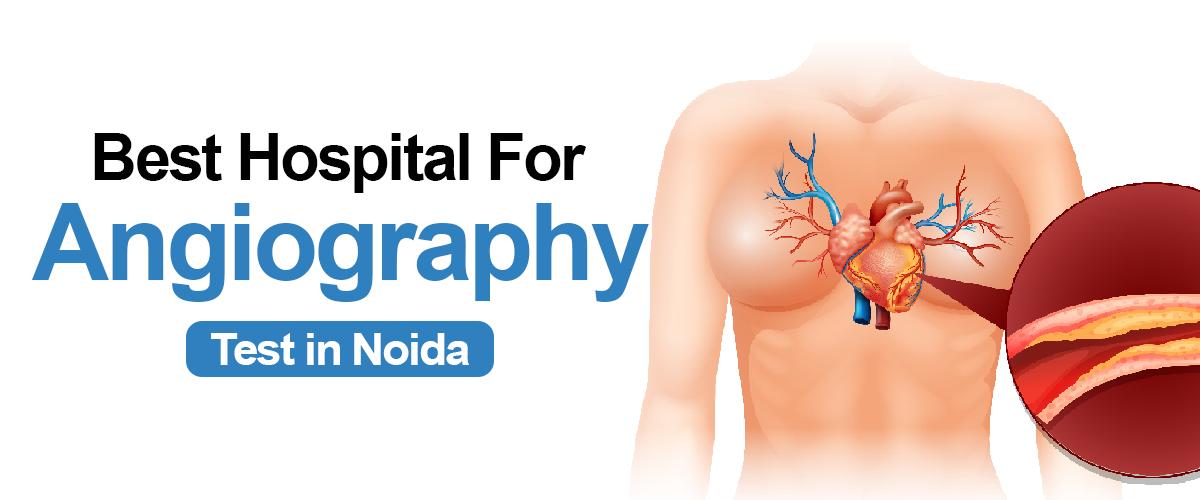
Angiography is a type of X-ray that helps doctors see your blood vessels. Blood vessels do not show clearly on a normal X-ray, so to make them visible, a special dye is injected into your blood. This dye highlights the blood vessels, and the X-ray images created during angiography are called angiograms. It is commonly performed to diagnose and evaluate conditions such as coronary artery disease, peripheral artery disease, and aneurysms. The angiography test time duration can differ based on various factors, such as the complexity of the procedure, the health condition of the patient, and the specific region being examined.
Book Your Appointment One click at +91 9667064100
Here are some key reasons why angiography is used:
Overall, angiography plays a crucial role in diagnosing, treating, and preventing various cardiovascular conditions, positively impacting patient care and promoting better outcomes.
Here is a step-by-step explanation of the process during angiography:
The angiography test time duration can last anywhere from half an hour to two hours. Generally, you will be allowed to return home in a few hours after finishing it.
Although angiography is generally regarded as a safe and efficient process, there are inherent dangers linked to any invasive medical procedure. These risks might differ depending on variables like the patient's overall well-being, the particular type of angiography being conducted, and the proficiency of the medical team involved. Several possible risks of angiography include:.
There exist various types of angiography, each specifically designed to capture visuals of particular blood vessels or organs.
There are three distinct approaches or methods for conducting angiography tests to detect blockages in blood vessels-.
CTA is less invasive as compared to traditional angiography. It utilizes advanced scanning technology to generate precise images of blood vessels in different areas of the body. To perform this procedure, a special dye is introduced into a vein via an IV line, and several CT scans are conducted while the dye flows through the bloodstream. The Computed Tomography angiography test time duration typically spans between 15 and 45 minutes.
Below are the essential steps to take before undergoing angiography:
After undergoing angiography, it is important to follow certain steps and take necessary precautions to guarantee the best possible recovery and reduce potential risks. Here is what you need to do after undergoing angiography:-
The price of the angiography examination in Noida, India, may differ based on different elements. The kind of angiography, the healthcare establishment or hospital where the test is taking place, and your insurance policy can all impact the price of the examination. Typically, the expense for angiography in Noida can fluctuate between INR 6,999 and INR 15,000. Nevertheless, the cost might increase in case supplementary procedures, like angioplasty or stenting, become necessary.
Simple non-invasive angiography procedures such as CT angiography may range from ₹8,000 to ₹15,000. Conversely, complex procedures like coronary angiography might cost between ₹20,000 to ₹1,50,000, depending on the chosen medical facility. But the angiography test cost in India can vary anywhere from ₹8,000 to ₹1,50,000 or higher, depending on the type and complexity of the procedure.
CT angiography is a type of medical examination that merges a CT scan with the infusion of a distinct coloring agent to produce visuals of blood vessels and tissues in a part of your body. The coloring agent is administered through an intravenous (IV) tube initiated in your forearm or hand. CT angiography is an essential diagnostic procedure that physicians may utilize to examine an individual's blood vessels. A CT angiography involves a doctor taking numerous X-rays of a person's body. In comparison to coronary angiography, CT angiography incorporates the utilization of multiple X-rays to assist the physician in generating a more detailed image of an individual's blood vessels. It also allows the physician to visualize the blood vessel structure of the patient in two or three dimensions. CT angiography is less invasive than coronary angiography and they pose fewer risks. Now, you will be pleased to learn that Felix Hospital offers the most economical price for CT angiography. The minimum CT angiography test near me can cost ₹ 10500.
We have a group of experienced cardiologists, radiologists, and technicians who excel in conducting angiography tests. We possess state-of-the-art equipment and modern facilities essential for performing the procedure in a safe and efficient manner. In the event of any complications during the angiography procedure, our medical facility is fully equipped to effectively manage emergencies. We have prompt availability to emergency revival tools, specialized medical staff, as well as intensive treatment wards, guaranteeing the well-being of patients at all moments. Searching for a CT angiography test near me will help in locating Felix Hospital. At Felix Hospital, we are committed to providing our patients with transparent and competitive pricing for the procedure compared to the angiography test cost in india.
Book Your Appointment on One click at +91 9667064100
Angiography is an important diagnostic test used to detect blockages or narrowing in blood vessels, especially in heart patients.

⚠️ Before You Read: The Most Important Thing on This Page
Most blogs — including many by reputed hospitals — falsely list bananas, papaya, spinach, okra, broccoli, and bitter gourd as "Vitamin D rich" foods. This is scientifically incorrect. This guide gives you the medically accurate, research-backed truth.
Get a quick Vitamin D test and a personalized bone health plan at Felix Hospital, Noida.
📞 Call Now- +91 96670 64100!
Vitamin D is the only vitamin your body can manufacture itself — your skin produces it when UVB rays from sunlight hit the skin. Yet despite India being one of the sunniest countries on Earth, we have one of the highest rates of Vitamin D deficiency in the world.
Technically, Vitamin D functions more like a steroid hormone than a traditional vitamin. Once synthesized or consumed, it gets converted in the liver to 25-hydroxyvitamin D [25(OH)D] — the storage form measured in blood tests — and then further activated in the kidneys to its active hormonal form, calcitriol.
In this active form, Vitamin D controls over 200 genes in the human body. It is essential for:
Bone Mineralization: Without Vitamin D, your intestines absorb only 10–15% of dietary calcium. With adequate Vitamin D, absorption rises to 30–40%. Deficiency causes rickets in children and osteomalacia or osteoporosis in adults.
Immune Regulation: Vitamin D activates T-cells and macrophages — the front-line soldiers of your immune system. Low levels increase susceptibility to respiratory infections, influenza, and autoimmune diseases like rheumatoid arthritis.
Muscle Function: Vitamin D receptors are present in muscle tissue. Deficiency causes proximal muscle weakness — making activities like climbing stairs or rising from chairs difficult, especially in older adults.
Cardiovascular Health: Studies link low Vitamin D to higher rates of hypertension, heart failure, and coronary artery disease. Vitamin D helps regulate the renin-angiotensin system that controls blood pressure.
Mental Health: Vitamin D receptors exist in the brain, and the vitamin in fluences serotonin synthesis. Deficiency correlates with depression, seasonal affective disorder (SAD), and cognitive decline.
Metabolic Health: Vitamin D improves insulin sensitivity and supports glucose metabolism. Deficiency is linked to higher risk of Type 2 diabetes — a critical concern for Indians, who have a genetic predisposition to metabolic disorders.
The following foods are commonly listed as "Vitamin D rich" by health websites, including those of established hospitals and insurance companies. All of these claims are factually incorrect based on USDA FoodData Central data and published nutritional science.
Food | What Sites Claim | Scientific Reality | Verdict |
Banana | "Rich in Vitamin D" | Contains 0 IU Vitamin D. Rich in magnesium which helps activate Vitamin D already present — completely different. | Myth |
Orange / Orange juice | "Good source of Vitamin D" | Natural oranges contain 0 IU. Only fortified OJ contains Vitamin D (~100 IU/cup). | Fortified Only |
Spinach (Palak) | "Contains Vitamin D" | Negligible Vitamin D (1–2 IU per 100g, essentially zero). Excellent source of calcium, iron, and magnesium — not Vitamin D. | Myth |
Broccoli | "Rich in Vitamin D" | Contains 0 IU Vitamin D. Rich in Vitamin K, C, and calcium — not Vitamin D. | Myth |
Okra (Bhindi) | "Fulfills Vitamin D needs" | Essentially 0 IU Vitamin D. No credible nutrition database lists okra as a Vitamin D source. | Myth |
Bitter Gourd (Karela) | "Houses Vitamin D" | Contains 0 IU Vitamin D. Useful for blood sugar management, not a Vitamin D source. | Myth |
Bottle Gourd (Lauki) | "Includes Vitamin D" | Contains 0 IU Vitamin D. Low-calorie vegetable with water content and fibre, not Vitamin D. | Myth |
Amla (Indian Gooseberry) | "Good amount of Vitamin D" | Contains 0 IU Vitamin D. One of the richest sources of Vitamin C — an entirely different nutrient. | Myth |
Pumpkin | "Contains Vitamin D" | Contains 0 IU Vitamin D. Rich in beta-carotene (Vitamin A precursor) and potassium. | Myth |
Kiwi / Papaya / Guava / Watermelon | "Vitamin D rich fruits" | All contain 0 IU Vitamin D. Excellent fruits for Vitamin C and antioxidants — not Vitamin D. | Myth |
Green Peas (Matar) | "Source of Vitamin D" | Contains 0 IU Vitamin D. Good for protein and fibre. | Myth |
Drumsticks (Moringa) | "Contains Vitamin D" | Contains 0 IU Vitamin D. Excellent source of calcium, iron, and Vitamin C. | Myth |
Mushrooms (dark-grown) | "Rich in Vitamin D" | Regular store mushrooms contain only 4–16 IU/85g. UV-exposed mushrooms are a real source. | Conditions Apply |
Many of these foods contain nutrients that work alongside Vitamin D — like magnesium (banana, spinach), calcium (moringa, kale), and Vitamin C (amla, guava). Poorly researched blogs confused these supportive nutrients with Vitamin D itself. The error then propagated across hundreds of websites.
Based on USDA FoodData Central, NIH Office of Dietary Supplements, and ICMR nutritional data, here are the actual foods that contain meaningful Vitamin D — with exact values:
Rank | Food | Serving | Vitamin D | Type | Best For |
1 | UV-Exposed Maitake Mushrooms | 85g | 2,000+ IU | Natural (D2) | Vegetarians / Vegans |
2 | Cod Liver Oil | 1 tablespoon | 1,360 IU | Natural (D3) | Supplement form |
3 | Rainbow Trout / Salmon (Rawas) | 85g cooked | 570–645 IU | Natural (D3) | Non-vegetarians |
4 | Swordfish / Surmai | 85g cooked | ~566 IU | Natural (D3) | Non-vegetarians |
5 | UV-Exposed Portobello Mushroom | 85g | ~400 IU | Natural (D2) | Vegetarians / Vegans |
6 | Mackerel (Bangda) | 85g cooked | ~360 IU | Natural (D3) | Non-vegetarians (coastal India) |
7 | UV-Exposed Shiitake (dried) | 85g | 100–1,600 IU | Natural (D2) | Vegetarians / Vegans |
8 | Canned Tuna (in water) | 85g | ~154 IU | Natural (D3) | Non-vegetarians |
9 | Fortified Soy Milk | 240ml | 107–117 IU | Fortified (D2/D3) | Vegetarians / Vegans |
10 | Fortified Cow's Milk | 240ml | 80–120 IU | Fortified (D3) | All (if brand is fortified) |
11 | Fortified Yogurt (Dahi) | 1 cup | 80–116 IU | Fortified | Vegetarians |
12 | Fortified Orange Juice | 240ml | ~100 IU | Fortified | All (if fortified — check label) |
13 | Whole Egg (yolk) | 1 large egg | 40–50 IU | Natural (D3) | Lacto-ovo vegetarians |
14 | Fortified Tofu | 100g | ~100 IU | Fortified | Vegetarians / Vegans |
15 | Sardines / Tarli (canned) | 2 pieces | ~46 IU | Natural (D3) | Affordable non-veg option |
16 | Beef Liver | 85g cooked | ~42 IU | Natural (D3) | Non-vegetarians |
17 | Fortified Breakfast Cereal | 1 serving (~30g) | 40–100 IU | Fortified | All (check label) |
18 | Cheese (Paneer / Processed) | 100g | 14–24 IU | Natural (small amount) | Vegetarians |
19 | Button Mushrooms (dark-grown, raw) | 85g | 4–16 IU | Natural (minimal) | Better than zero |
Mushrooms occupy a unique biological position: they are fungi (not plants, fruits, or vegetables), yet they are the only non-animal food that naturally produces Vitamin D. Here is everything you need to know to use them effectively in India.
Mushrooms contain ergosterol — a sterol compound in their cell membranes. When UVB rays from sunlight (or a UV lamp) hit ergosterol, it converts to ergocalciferol, which is Vitamin D2. This process mirrors how human skin converts cholesterol to Vitamin D3 under sunlight.
Mushroom Type | Vitamin D (per 85g) | Notes |
Regular button mushrooms (dark-grown, store-bought) | 4–16 IU | Most common in India; very low Vitamin D |
Button mushrooms — sun-treated 30 min (gill-side up) | ~400 IU | DIY method — very effective |
Wild maitake mushrooms (UV-exposed) | 2,000+ IU | Rare in India; available at specialty stores |
Shiitake (dried, UV-exposed) | 100–1,600 IU | Available online and specialty health stores |
Portobello (gill-side up, 1 hr direct sun) | ~400–1,000 IU | Available in metro cities; easy DIY sun treatment |
Oyster mushrooms (UV-treated) | ~200–400 IU | Increasingly common across India |
Slice or open: Slice mushrooms or place whole ones gill-side up on a tray.
Time it right: Place in direct sunlight between 11 AM and 2 PM.
Wait 30–60 minutes: Minimum 30 minutes; 60 minutes doubles the effect.
Cook normally: Vitamin D is heat-stable — cooking does not destroy it.
Store for later: Sun-treated dried mushrooms retain Vitamin D for months.
Important: UVB rays do not pass through glass. The mushrooms must be in outdoor direct sunlight, not on a windowsill inside.
Fortified Product | Vitamin D per Serving | What to Check on Label | Indian Brands (Examples) |
Milk (Fortified) | 80–120 IU/cup | Look for "Vitamin D3" or "Cholecalciferol" in ingredients | Amul Taaza, Mother Dairy (some variants — verify label) |
Soy Milk | 107–117 IU/cup | "Fortified with Vitamin D" | Sofit, Epigamia plant milk |
Dahi / Yogurt (Fortified) | 80–116 IU/cup | Check label specifically; most Indian dahi is NOT fortified | Epigamia Greek yogurt (some variants) |
Tofu (Fortified) | ~100 IU/100g | "Fortified tofu" or Vitamin D in nutrition table | Soy India, imported brands at health stores |
Breakfast Cereals | 40–100 IU/serving | "Fortified with Vitamin D" | Kellogg's (check variant), Quaker (some) |
Orange Juice (Fortified) | ~100 IU/cup | Most Indian juices are NOT fortified — check label | Some imported brands; fresh OJ = 0 IU |
India does not mandate Vitamin D fortification of staple foods the way the US, UK, and Canada do. Do not assume any product is fortified — check the nutrition facts table specifically for "Vitamin D" and the ingredients list for "Cholecalciferol" (D3) or "Ergocalciferol" (D2). If neither appears, the product contains no meaningful Vitamin D.
The following Indian fruits and vegetables do not contain Vitamin D, but they play a genuine supporting role in Vitamin D metabolism.
# | Food | How It Supports Vitamin D | Key Nutrient | Serving Tip |
1 | Banana | Magnesium activates Vitamin D in liver and kidneys | Magnesium (32 mg/banana) | 1–2 per day |
2 | Avocado | Healthy fats improve Vitamin D absorption (fat-soluble) | Monounsaturated fats | Half with a Vitamin D-rich meal |
3 | Spinach (Palak) | Magnesium + calcium synergy with Vitamin D for bone health | Magnesium, Calcium | 1 cup cooked; saag or dal palak |
4 | Amla (Indian Gooseberry) | High Vitamin C reduces oxidative stress affecting Vitamin D receptors | Vitamin C (600+ mg/100g) | 1 fresh or juice daily |
5 | Moringa / Drumstick Leaves | Calcium + magnesium support bone matrix that Vitamin D mineralizes | Calcium (200 mg/100g), Mg | Add to dal or soup |
6 | Fenugreek (Methi) Leaves | Magnesium source; supports Vitamin D activation pathway | Magnesium, Iron | Methi paratha, sabzi |
7 | Kale | High calcium works synergistically with Vitamin D for bone formation | Calcium (150 mg/100g) | Salads, smoothies |
8 | Amaranth Leaves (Chaulai) | Calcium and magnesium support; good iron for overall bone health | Calcium, Iron, Mg | Saag; stir fry |
9 | Sweet Potato | Magnesium content; beta-carotene supports immune function | Magnesium, Vitamin A | Boiled or baked (skin on) |
10 | Pumpkin Seeds (Kaddu Ke Beej) | One of the richest plant sources of magnesium (150 mg/28g) | Magnesium | 28g as daily snack |
11 | Dark Chocolate (70%+) | Magnesium (64 mg/28g) supports Vitamin D activation | Magnesium | 1–2 squares daily |
12 | Almonds (Badam) | Magnesium + healthy fats for fat-soluble Vitamin D absorption | Magnesium, Fats | 10–12 soaked almonds daily |
13 | Papaya | Vitamin C and antioxidants support immune function alongside Vitamin D | Vitamin C, Folate | 1 cup for breakfast |
14 | Guava | Vitamin C and fibre; immune support companion to Vitamin D | Vitamin C (228 mg/100g) | 1 whole guava as snack |
15 | Kiwi | Vitamin C and K; supports collagen and bone matrix health | Vitamin C, Vitamin K | 2 kiwis as snack |
16 | Bitter Gourd (Karela) | Improves insulin sensitivity — linked to better Vitamin D utilization | Chromium, Iron | 2x weekly in sabzi |
17 | Broccoli | Vitamin K2 supports how Vitamin D directs calcium to bones (not arteries) | Vitamin K, Calcium | Half cup steamed |
18 | Cauliflower | Vitamin K and glucosinolates support bone health pathways | Vitamin K, Vitamin C | Roasted, curried |
19 | Fig (Anjeer) | Calcium and phosphorus build the bone matrix Vitamin D mineralizes | Calcium, Phosphorus | 2–3 dried figs daily |
20 | Oranges (fresh) | Vitamin C protects Vitamin D receptors from oxidative damage | Vitamin C | 1 orange or 1 glass fresh juice |
21 | Watermelon | Lycopene reduces inflammation; hydration supports kidney health | Lycopene, water | 2 cups in summer |
22 | Bottle Gourd (Lauki) | Supports kidney health — kidneys activate Vitamin D to its hormonal form | Water, potassium | Lauki ki sabzi or juice |
23 | Ridge Gourd (Turai) | Low calorie, supports metabolic health linked to Vitamin D regulation | Fibre, Vitamins B, C | Light sabzi |
24 | Green Peas (Matar) | Magnesium and Vitamin K support bone health complementary to Vitamin D | Magnesium, Vit K | Add to rice, curries |
25 | Pineapple | Bromelain enzyme reduces inflammation; Vitamin C supports immunity | Vitamin C, Bromelain | Half cup fresh; avoid canned |
Being vegetarian in India and maintaining optimal Vitamin D is genuinely challenging — but not impossible. Here is the complete strategy, ranked by effectiveness:
Priority 1 — Sun-Treated Mushrooms (3–4x per week): The single most impactful dietary change a vegetarian can make. Sun-treat button or oyster mushrooms for 30–60 minutes before cooking and consume 85–100g per serving. This alone can provide 300–400+ IU per serving.
Priority 2 — 2 Whole Eggs Daily (lacto-ovo vegetarians): Two egg yolks provide 80–100 IU of naturally occurring Vitamin D3 — the most bioavailable plant-adjacent source. Pasture-raised or desi eggs have higher Vitamin D due to the hen's sun exposure.
Priority 3 — Fortified Dairy Daily: Switch to fortified milk or yogurt as your daily dairy choice. One cup of fortified milk plus one serving of fortified yogurt can add 160–236 IU to your daily total.
Priority 4 — Magnesium-Rich Foods (Daily): Ensure adequate magnesium so your body can activate the Vitamin D you get. Daily banana + a handful of pumpkin seeds or almonds + spinach-based sabzi covers your magnesium needs effectively.
Priority 5 — Vitamin D3 Supplement (Under Doctor Guidance): Given the reality of Indian vegetarian diets, a Vitamin D3 supplement (from lichen — the vegan form) is often the most practical solution after a blood test confirms deficiency. Do not self-dose — get tested first.
Day 1 — Monday (Vegetarian: approximately 450 IU from food + sun exposure)
Breakfast: Fortified oats with fortified soy milk + 2 boiled eggs + 1 kiwi — approximately 180 IU from eggs + milk
Lunch: Sun-treated mushroom and spinach sabzi + 2 roti + 1 cup fortified dahi — approximately 480 IU from mushrooms + dahi
Snack: 1 banana + 10 almonds + 1 orange — magnesium boost for Vitamin D activation
Dinner: Moringa dal + cauliflower sabzi + 1 cup fortified milk before bed — approximately 100 IU from milk
Day 2 — Tuesday (Non-Vegetarian: approximately 700 IU from food)
Breakfast: 2-egg omelette cooked in olive oil + fortified milk chai + 1 banana — approximately 100 IU from eggs + milk
Lunch: Grilled Bangda (mackerel) fish + steamed rice + palak dal + salad — approximately 360 IU from mackerel alone
Snack: 1 cup fortified dahi with amla chutney — approximately 100 IU + Vitamin C boost
Dinner: Chicken liver curry + roti + kachumber salad with kiwi — approximately 40 IU + iron and B12
Day 3 to 7 — Principle-Based Rotation
Core Rule: Include at least one of the following every single day — sun-treated mushrooms, oily fish (bangda/rawas/surmai),or 2 eggs. Rotate between them and pair with fortified milk or yogurt daily.
Magnesium: Include spinach, banana, or almonds daily to ensure Vitamin D activation. Deficiency in magnesium renders even supplemental Vitamin D less effective.
Fat Pairing: Always consume Vitamin D-containing foods with a fat source — ghee, coconut oil, avocado, or nuts. Vitamin D is fat-soluble and absorption increases 32% with dietary fat.
India receives more annual sunlight than almost any European country. Yet our deficiency rates are dramatically higher. This paradox has a scientific explanation — and it means most Indians are not getting effective Vitamin D from sun exposure.
Air Pollution (Delhi/Noida): Particulate matter (PM2.5) in cities like Noida, Delhi, and Gurugram scatters and absorbs UVB radiation before it reaches your skin. Studies show urban Indians in North India receive 50–70% less effective UVB compared to rural areas — even on clear days.
Window Glass Blocks UVB: Standard glass completely blocks UVB rays. If you are sitting in a sunny office or car, you are getting zero Vitamin D synthesis. Only direct outdoor sun works.
Skin Pigmentation: Melanin — the pigment giving darker skin tones — acts as natural sunscreen. Indians need 3–5 times more sun exposure than someone with light skin to produce the same amount of Vitamin D.
Clothing Coverage: Full-sleeve clothing, dupattas, and other cultural garments block UVB exposure. Effective Vitamin D synthesis requires exposed skin — arms and legs at minimum.
Wrong Time of Day: UVB rays are only strong enough for Vitamin D synthesis when the sun is high in the sky, roughly 11 AM to 2 PM IST. Morning walks and evening strolls, while beneficial for health, produce little Vitamin D.
Indoor Lifestyle: Office workers, students, and urban Indians typically spend 90% or more of daylight hours indoors. Air conditioning and screen culture have dramatically reduced our sun contact compared to previous generations.
Time: 11 AM to 2 PM IST — UVB intensity is sufficient only during this window
Duration: 20–30 minutes for lighter skin tones; 40–60 minutes for darker skin tones
Exposed area: Arms and legs must be exposed — face and hands alone are insufficient surface area
Frequency: At least 3–4 times per week
Location: Must be outdoors in direct sunlight, not behind glass
Sunscreen timing: Apply sunscreen after 20–30 minutes of sun exposure — not before, as sunscreen blocks UVB
Season: In North India (Delhi, Noida), Vitamin D synthesis from sunlight is significantly reduced from November to February
Deficiency Risk Checklist:
Check any symptoms you have experienced in the past 3 months:
Unexplained bone pain or aching joints, especially in the lower back, hips, or legs
Persistent fatigue or low energy despite adequate sleep (7–8 hours)
Muscle weakness — difficulty climbing stairs, getting up from a chair, or carrying groceries
Getting sick frequently — repeated colds, respiratory infections, or slow recovery
Low mood, depression, or unexplained anxiety (especially in winter months)
Hair loss or slow hair growth
Slow wound healing
Bone fractures from minor injuries or falls
Child with bowed legs, delayed walking, or dental problems (possible rickets)
Spending most of the day indoors with minimal sun exposure
If you checked 3 or more items, we strongly recommend getting a serum 25(OH)D blood test. This is a simple blood test available at most diagnostic labs in India for Rs. 500–1,200.
High-Risk Group | Why | Deficiency Prevalence |
Postmenopausal women | Estrogen loss reduces Vitamin D receptor sensitivity; reduced outdoor activity | 80–90% |
Office workers (urban) | Indoor lifestyle; no meaningful sun exposure during working hours | 70–85% |
Elderly (65+) | Skin produces 75% less Vitamin D than young skin; reduced kidney activation | 80–90% |
Pregnant women | Fetal demand; hormonal changes affecting Vitamin D metabolism | 70–80% |
Obese individuals | Vitamin D is fat-soluble; stored in body fat and unavailable for use | 75–85% |
Vegetarians / Vegans | No fatty fish or egg yolk consumption; limited dietary sources | 65–80% |
Individuals with diabetes | Kidney dysfunction impairs Vitamin D activation; metabolic dysregulation | 75–90% |
Serum 25(OH)D Level | Classification | What This Means | Action |
Above 100 ng/mL | Toxicity Risk | Hypercalcemia risk; excess supplementation | Stop supplements; see doctor immediately |
30–100 ng/mL | Optimal / Normal | Sufficient for bone health and overall function | Maintain through diet and sun |
20–29 ng/mL | Insufficient | Suboptimal; increased disease risk | Increase dietary sources + supervised supplement |
12–19 ng/mL | Deficient | Bone loss, immune impairment, muscle weakness | Medical treatment required; high-dose supplementation |
Below 12 ng/mL | Severely Deficient | Rickets risk in children; osteomalacia in adults | Urgent medical treatment; possible IV/injection |
Group | Recommended Daily Intake | Tolerable Upper Limit | Common Prescription in India |
Infants 0–12 months | 400 IU (10 mcg) | 1,000–1,500 IU | Drops: 400 IU/day |
Children 1–18 years | 600 IU (15 mcg) | 2,500–4,000 IU | 60,000 IU sachet every 2 weeks (if deficient) |
Adults 19–70 years | 600 IU (15 mcg) | 4,000 IU | 60,000 IU sachet weekly x 8–12 weeks; then 1,000–2,000 IU/day maintenance |
Adults 70+ years | 800 IU (20 mcg) | 4,000 IU | Same as adults; often continued longer |
Pregnant women | 600 IU (15 mcg) | 4,000 IU | As prescribed by OB-GYN based on blood test |
Breastfeeding women | 600 IU (15 mcg) | 4,000 IU | Supplement baby separately if breastfed exclusively |
People with deficiency (below 20 ng/mL) | Doctor-prescribed | As advised | Loading dose 60,000 IU/week x 8–12 weeks; test and adjust |
Vitamin D toxicity (hypervitaminosis D) is possible with excessive supplementation. Symptoms include nausea, vomiting, weakness, confusion, excessive thirst, kidney stones, and in severe cases, heart arrhythmias. The upper safe limit is 4,000 IU/day for adults. Therapeutic high doses prescribed by doctors are safe for limited periods because they are intermittent. Do not self-prescribe high-dose supplements.
Experiencing any of the symptoms above? Don't guess your Vitamin D levels. Book a test at Felix Hospital today: +91 96670 64100.
Office of Dietary Supplements, NIH. Vitamin D: Fact Sheet for Health Professionals. Updated 2025. ods.od.nih.gov
Japelt RB, Jakobsen J. Vitamin D in plants: A review of occurrence, analysis, and biosynthesis. Frontiers in Plant Science. 2013;4:136.
Haytowitz DB. Vitamin D in mushrooms. USDA Nutrient Data Laboratory, Agricultural Research Service. 2009.
Keegan RJH, et al. Photobiology of vitamin D in mushrooms and its bioavailability in humans. Dermato-Endocrinology. 2013;5(1):165–176.
USDA FoodData Central. Nutritional database for standard reference. fdc.nal.usda.gov
Dietary Guidelines for Americans 2020–2025. Food Sources of Vitamin D. dietaryguidelines.gov
ICMR-NIN. Nutrient Requirements and Recommended Dietary Allowances for Indians. Indian Council of Medical Research. 2020.
Harinarayan CV, Joshi SR. Vitamin D status in India — Its implications and remedial measures. Journal of Association of Physicians of India. 2009;57:40–48.
Aparna P, et al. Low dietary calcium intake and prevalence of vitamin D deficiency in South Asia. Asia Pacific Journal of Clinical Nutrition. 2015.
Black LJ, et al. In pursuit of vitamin D in plants. Nutrients. 2017;9(2):136.
Dr. Ravi Sharma is a senior physician with 42+ years of experience, known for compassionate and comprehensive patient care in Noida.

Protein powder supplements are a rage now. They sure have benefits – they help the body to meet its daily protein needs. And that too, in a convenient way.
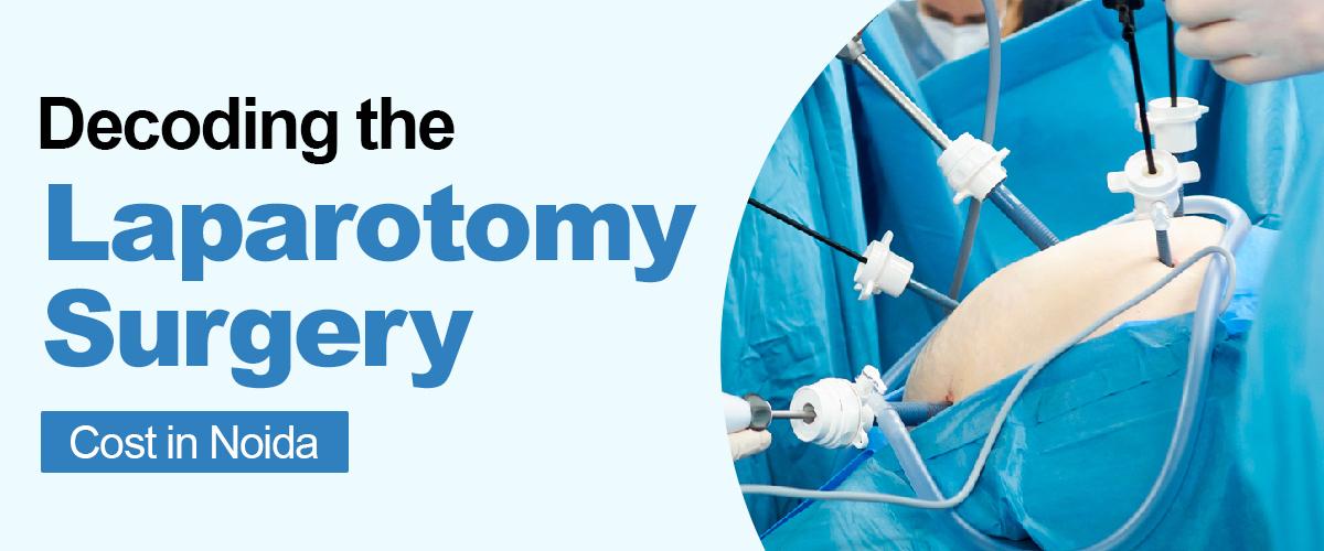
Expert gastroenterology care for better digestive health, offering advanced diagnosis and treatment for GERD, IBS, ulcers, and liver disorders.
Understand the factors that influence gastric balloon surgery cost in Noida, including hospital facilities, doctor expertise, and post-procedure care.

A complete guide to understanding varicose vein surgery expenses in Noida, including treatment types, hospital charges, and recovery costs.
Appendix surgery, also known as appendectomy, is a common emergency procedure performed to treat appendicitis.

Subscribe to our
© Copyright 2026. All Rights Reserved by Felix Healthcare Private Limited
Design and development by :


 +(91)9667064100
 Emergency : +(91)9667064100