 WhatsApp
 WhatsApp


Subscribe to our
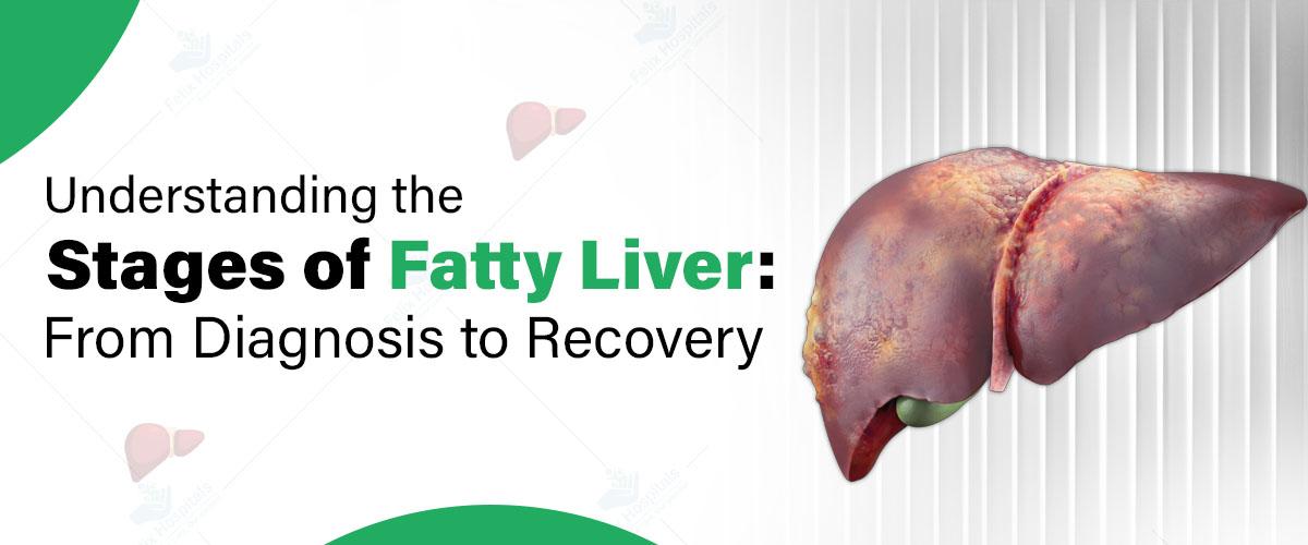
Fatty liver disease is a growing health concern worldwide, affecting millions of individuals regardless of age or lifestyle. This condition arises when excess fat accumulates in the liver cells, potentially leading to severe health complications if left unmanaged. Early detection and effective management are crucial for reversing the condition and preventing progression to advanced stages. Consulting the best gastroenterologist hospital in Noida can ensure timely diagnosis and personalized care for managing fatty liver disease. This blog aims to provide an in-depth understanding of fatty liver disease, covering its stages, diagnosis methods, treatment strategies, and tips for long-term management.
If you or someone you know may be at risk, consult a healthcare professional today. Call +(91)9667064100 or click here for more information.
Fatty liver disease refers to the accumulation of fat in liver cells, which can interfere with normal liver function. It is broadly classified into two types:
Alcoholic Fatty Liver Disease (AFLD): Caused by excessive alcohol consumption.
Non-Alcoholic Fatty Liver Disease (NAFLD): This occurs in individuals who consume little to no alcohol and is commonly linked to obesity, diabetes, and metabolic syndrome.
Factors contributing to fatty liver disease are multifaceted and often interrelated. These include:
Obesity and Sedentary Lifestyle: Excess body weight, particularly abdominal fat, is a significant risk factor. A sedentary lifestyle exacerbates fat accumulation in the liver.
Type 2 Diabetes and Insulin Resistance: Insulin resistance promotes fat storage in the liver, increasing the likelihood of developing NAFLD.
High Cholesterol and Triglycerides: Elevated levels of these lipids can contribute to fat buildup and inflammation in the liver.
Excessive Alcohol Consumption: Chronic alcohol intake is the primary cause of AFLD, impairing the liver's ability to metabolize fats.
Genetic Predisposition: Certain genetic factors make some individuals more susceptible to developing fatty liver disease.
Understanding these risk factors is crucial for both prevention and early intervention.
Fatty liver disease progresses through four distinct stages:
At this initial stage, fat accumulates in the liver cells without causing inflammation or damage. Simple fatty liver is often asymptomatic and may only be detected through imaging studies or routine health check-ups.
This stage involves inflammation and damage to liver cells. NASH is a more severe form of fatty liver disease and can progress to fibrosis if not addressed. Early symptoms of fatty liver may include fatigue, abdominal discomfort, and mild liver enlargement.
In fibrosis, persistent inflammation leads to the formation of scar tissue around the liver cells. While the liver may still function, its efficiency is reduced, and further progression increases the risk of complications.
Cirrhosis represents the most advanced stage, characterized by extensive scarring and irreversible damage. Complications such as liver failure, portal hypertension, and liver cancer can arise at this stage, requiring advanced medical interventions.
Early diagnosis is key to preventing the progression of fatty liver disease. Common diagnostic steps include:
Fatigue and weakness
Pain or discomfort in the upper right abdomen
Unexplained weight loss
Blood Tests: Elevated liver enzymes (ALT, AST) may indicate liver inflammation.
Imaging Studies: Ultrasound, CT, or MRI scans help visualize fat accumulation and structural changes in the liver.
Routine check-ups are especially important for individuals with risk factors such as obesity or diabetes.
Fatty liver disease is often reversible with timely intervention and lifestyle modifications. Treatment strategies include:
Healthy Diet: Focus on whole grains, lean proteins, fresh fruits, and vegetables while avoiding high-fat and high-sugar foods.
Regular Physical Activity: Engage in at least 30 minutes of moderate exercise most days of the week.
Medications to manage underlying conditions such as diabetes, cholesterol, or hypertension.
Regular monitoring of liver health to assess treatment progress.
For AFLD, reducing or eliminating alcohol consumption is critical to prevent further liver damage.
Weight loss of 5-10% of body weight can significantly improve liver health and reduce fat accumulation.
In severe cases, options such as liver transplantation may be necessary to manage complications arising from cirrhosis or liver failure.
Preventing fatty liver disease and ensuring long-term management involves:
Balanced Diet: Prioritize nutrient-rich foods and maintain portion control.
Regular Exercise: Incorporate activities like walking, swimming, or yoga into your daily routine.
Hydration: Drink adequate water throughout the day.
Stress Management: Practice mindfulness, meditation, or other relaxation techniques.
Routine Screenings: Regularly check liver health, especially if you have risk factors.
At Felix Hospitals, we are proud to introduce our esteemed team of gastroenterologists, dedicated to delivering exceptional care and expertise for all your digestive health concerns.
Don’t wait for symptoms to worsen, Consult a healthcare professional today to start your journey toward recovery. Call now at +91 9667064100.
Fatty liver disease is a progressive condition that can have severe consequences if ignored. Understanding the stages of fatty liver, recognizing early symptoms, and implementing effective lifestyle changes are vital to reversing the disease and maintaining a healthy liver. With timely medical intervention from an experienced Liver specialist in Noida and a proactive approach, recovery and long-term liver health are achievable. Consulting the right expert ensures accurate diagnosis, personalized treatment, and guidance for sustaining overall liver wellness.
Q1. How can I differentiate between the symptoms of NAFLD and other liver-related conditions?
Ans: While fatigue and abdominal discomfort are common to many liver conditions, NAFLD is often asymptomatic in the early stages of fatty liver and may only be identified through routine tests. Advanced symptoms like significant weight loss or yellowing of the skin (jaundice) might indicate more severe liver issues, warranting further investigation.
Q2. Is fatty liver disease reversible at any stage, or only in the early phases?
Ans: Fatty liver disease is most reversible during the early stages, such as simple fatty liver (steatosis). Once the condition progresses to cirrhosis, the damage is often permanent, but management strategies can slow further deterioration.
Q3. Can individuals with a healthy weight still develop fatty liver disease?
Ans: Yes, individuals with a healthy weight can develop fatty liver disease, especially if they have other risk factors such as type 2 diabetes, high cholesterol, or genetic predisposition. This condition is termed lean NAFLD.
Q4. How often should at-risk individuals undergo liver function tests or imaging?
Ans: At-risk individuals, such as those with obesity, diabetes, or a family history of liver disease, should undergo liver function tests annually or as recommended by their healthcare provider. Imaging tests like ultrasound may be suggested every couple of years for monitoring.
Q5. Does fatty liver disease increase the risk of other health conditions?
Ans: Yes, fatty liver disease can increase the risk of cardiovascular diseases, type 2 diabetes, and chronic kidney disease. Advanced stages, such as cirrhosis, can also lead to liver cancer.
Understand the factors that influence gastric balloon surgery cost in Noida, including hospital facilities, doctor expertise, and post-procedure care.

Expert gastroenterology care for better digestive health, offering advanced diagnosis and treatment for GERD, IBS, ulcers, and liver disorders.
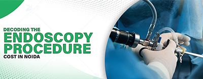
Understand the factors that influence endoscopy procedure cost in Noida. Learn about the types, preparation, and benefits of choosing the best multi-speciality hospital in Noida for accurate diagnosis.
Appendix surgery, also known as appendectomy, is a common emergency procedure performed to treat appendicitis.

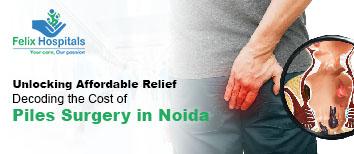
बवासीर, जिसे पाइल्स या हेमोराइड्स के नाम से भी जाना जाता है, एक आम समस्या है, जो हर उम्र के लोगों को प्रभावित कर सकती है। इसमें मलाशय (रेक्टम) के आसपास या अंदर की नसों में सूजन हो जाती है, जिससे दर्द, खुजली और कभी-कभी रक्तस्राव जैसी परेशानियाँ होती हैं। अगर समय पर सही इलाज न मिले, तो यह समस्या गंभीर हो सकती है और ऑपरेशन की जरूरत पड़ सकती है। अगर आप बवासीर से परेशान हैं और ऑपरेशन के विकल्प पर विचार कर रहे हैं, तो यह ब्लॉग आपकी मदद कर सकता है। इसमें हम बवासीर के कारणों, इसके ऑपरेशन के फायदे-नुकसान और साथ ही नोएडा के सर्वश्रेष्ठ हॉस्पिटल में उपचार के बारे में जानेंगे।
क्या आपको भी बवासीर से निजात चाहिए? आज ही फ़ेलिक्स हॉस्पिटल में अपॉइंटमेंट बुक करें और विशेषज्ञ से परामर्श लें। हमसे संपर्क करें +91 9667064100.
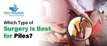
बवासीर (Hemorrhoids) मलाशय और गुदा (Anus) के आसपास की नसों में सूजन और दर्द का कारण बनने वाली एक स्थिति है। इसमें अक्सर गुदा के अंदर या बाहर गांठ या मस्से बन जाते हैं। यह स्थिति सामान्यतः दो प्रकार की होती है:
आंतरिक बवासीर - ये गुदा के अंदर होती है और मल त्यागते समय रक्तस्राव हो सकता है।
बाहरी बवासीर - ये गुदा के बाहर होती है, जिसमें दर्द और खुजली होती है।
बवासीर के कई कारण होते हैं, जो जीवनशैली, आहार, और स्वास्थ्य संबंधित आदतों से जुड़े हो सकते हैं। यह समस्या विशेष रूप से तब उभरती है जब गुदा और मलाशय के आसपास की नसों पर अत्यधिक दबाव पड़ता है। इसके कारण निम्नलिखित हैं:
कब्ज़ - लंबे समय तक कब्ज़ रहना बवासीर का सबसे आम कारण है। जब व्यक्ति कब्ज़ की वजह से बार-बार जोर लगाकर मल त्याग करता है, तो गुदा के आसपास की नसों पर दबाव बढ़ता है, जिससे ये सूज जाती हैं और बवासीर की समस्या उत्पन्न होती है।
लंबे समय तक बैठे रहना - अत्यधिक समय तक एक ही स्थिति में बैठे रहने से भी बवासीर का खतरा बढ़ता है। विशेषकर ऑफिस में लंबे समय तक बैठे रहना, बिना किसी गतिविधि के दिन बिताना गुदा और मलाशय की नसों पर दबाव डाल सकता है। इस तरह के दबाव से नसों में सूजन हो सकती है और बवासीर हो सकता है।
अधिक वजन - मोटापा भी बवासीर का एक प्रमुख कारण है। अधिक वजन होने पर पेट और गुदा की नसों पर अतिरिक्त दबाव पड़ता है, जिससे इन नसों में सूजन आ सकती है और बवासीर का खतरा बढ़ जाता है।
गर्भावस्था - गर्भवती महिलाओं को बवासीर की समस्या अधिक होती है। गर्भावस्था में बढ़ते बच्चे के कारण गुदा और पेट की नसों पर दबाव बढ़ जाता है। इसके अलावा, हार्मोनल बदलाव के कारण भी गर्भवती महिलाओं में बवासीर की संभावना बढ़ सकती है।
अनियमित और असंतुलित आहार - ज्यादा तला हुआ, मसालेदार, और कम फाइबर युक्त भोजन का सेवन करने से भी बवासीर की समस्या हो सकती है। कम फाइबर के कारण मल त्याग में कठिनाई होती है, जिससे कब्ज़ और बवासीर का खतरा बढ़ जाता है। फाइबर युक्त भोजन से मल को नरम रखने में मदद मिलती है और इससे बवासीर की संभावना घटती है।
बवासीर का ऑपरेशन तब आवश्यक हो सकता है जब स्थिति इतनी गंभीर हो जाए कि अन्य उपचार काम न करें और दर्द असहनीय हो। इसके कुछ प्रमुख संकेत निम्नलिखित हैं:
असहनीय दर्द - बवासीर की स्थिति कभी-कभी इतनी बिगड़ जाती है कि व्यक्ति को दिन-रात असहनीय दर्द का सामना करना पड़ता है। यह दर्द दैनिक जीवन की गतिविधियों में रुकावट डाल सकता है और व्यक्ति को सामान्य कार्यों में कठिनाई महसूस हो सकती है। ऐसी स्थिति में ऑपरेशन एक कारगर विकल्प हो सकता है।
अत्यधिक रक्तस्राव - अगर मल त्यागते समय लगातार अत्यधिक मात्रा में रक्तस्राव हो रहा है, तो यह एक गंभीर स्थिति हो सकती है। लगातार रक्तस्राव से व्यक्ति को एनीमिया होने का खतरा बढ़ जाता है। ऐसे में बवासीर का ऑपरेशन करना आवश्यक हो सकता है।
अन्य उपचार विफल हो जाएं - कई बार दवाइयों, मलहम, और गैर-शल्य चिकित्सा उपचार जैसे बैंडिंग या स्क्लेरोथेरेपी का असर बवासीर पर नहीं होता है। यदि सभी सामान्य उपाय बेअसर साबित हो रहे हैं, तो ऑपरेशन आवश्यक हो सकता है।
गांठ का आकार बढ़ना - जब बवासीर के मस्से या गांठें इतनी बड़ी हो जाएं कि वे अपने आप अंदर न जाएं या मल त्याग में बाधा उत्पन्न करें, तब ऑपरेशन की जरूरत पड़ सकती है। बड़े मस्से व्यक्ति को असहनीय असुविधा और दर्द का कारण बन सकते हैं।
बवासीर के ऑपरेशन से कई लाभ हो सकते हैं, जो मरीज़ के स्वास्थ्य और जीवन की गुणवत्ता को बेहतर बनाते हैं। ऑपरेशन के कुछ प्रमुख फायदे इस प्रकार हैं:
दर्द से राहत - बवासीर का ऑपरेशन कराने के बाद दर्द से काफी हद तक राहत मिलती है। जो व्यक्ति लंबे समय से दर्द झेल रहे होते हैं, उन्हें ऑपरेशन के बाद तुरंत आराम मिलता है।
रक्तस्राव में कमी - ऑपरेशन के बाद मल त्यागते समय होने वाले रक्तस्राव में भी काफी कमी आती है। इससे व्यक्ति एनीमिया जैसी समस्याओं से बच सकता है और बेहतर स्वास्थ्य का अनुभव कर सकता है।
जीवन की गुणवत्ता में सुधार - बवासीर का ऑपरेशन कराने से व्यक्ति की जीवन की गुणवत्ता में सुधार आता है। वह सामान्य जीवन जी पाता है और अपनी दैनिक गतिविधियाँ बिना किसी असुविधा के कर पाता है।
स्थायी समाधान - ऑपरेशन के बाद बवासीर की समस्या से स्थायी राहत मिलती है और भविष्य में इसके पुनः उत्पन्न होने की संभावना कम हो जाती है। ऐसे में यह एक दीर्घकालिक समाधान साबित हो सकता है।
हर सर्जरी की तरह, बवासीर के ऑपरेशन में भी कुछ संभावित जोखिम होते हैं, जिनमें शामिल हैं:
संक्रमण का खतरा - ऑपरेशन के बाद संक्रमण का खतरा हो सकता है। हालांकि, एंटीबायोटिक्स के उपयोग से इस खतरे को नियंत्रित किया जा सकता है, लेकिन संक्रमण की थोड़ी संभावना बनी रहती है।
दर्द और सूजन - ऑपरेशन के बाद कुछ समय तक दर्द और सूजन की समस्या हो सकती है। हालांकि, समय के साथ यह ठीक हो जाती है, लेकिन इसके लिए धैर्य और सावधानी की आवश्यकता होती है।
रक्तस्राव - कुछ मामलों में ऑपरेशन के बाद भी रक्तस्राव हो सकता है। यह स्थिति दुर्लभ होती है, लेकिन इसकी संभावना को नज़रअंदाज नहीं किया जा सकता।
अन्य जटिलताएँ - कभी-कभी ऑपरेशन के बाद मल त्याग में कठिनाई, अस्थायी कब्ज़, या गुदा की कमजोरी जैसी समस्याएँ भी उत्पन्न हो सकती हैं।
फ़ेलिक्स हॉस्पिटल में बवासीर के इलाज के लिए बेहतरीन सुविधा और अनुभवी चिकित्सक उपलब्ध हैं। यहां के विशेषज्ञ, डॉ. रितेश अग्रवाल, नोएडा के कुशल जनरल सर्जन हैं और बवासीर के ऑपरेशन में महारत रखते हैं। उनके मार्गदर्शन में बवासीर का इलाज सटीकता और सावधानी से किया जाता है, जिससे मरीज़ को जल्दी और स्थायी राहत मिलती है।
अनुभवी डॉक्टरों की टीम
अत्याधुनिक उपकरण और तकनीक
रोगी के प्रति संवेदनशील और सहायक स्टाफ
सर्जरी के बाद मरीज़ की देखभाल और फॉलो-अप के लिए विशेष इंतजाम
अगर आप बवासीर के कारण असुविधा का सामना कर रहे हैं, तो संकोच न करें। फ़ेलिक्स हॉस्पिटल में एक बेहतरीन उपचार प्राप्त करने के लिए यहां क्लिक करें।
बवासीर एक सामान्य लेकिन दर्दनाक स्थिति है, जो कई बार ऑपरेशन तक ले जा सकती है। हालाँकि, सही समय पर इलाज और उचित देखभाल से इसे नियंत्रित किया जा सकता है। बवासीर के ऑपरेशन के फायदे और नुकसान को ध्यान में रखते हुए, सही निर्णय लेना आवश्यक है। फ़ेलिक्स हॉस्पिटल के विशेषज्ञ डॉक्टरों और नवीनतम चिकित्सा सुविधाओं के साथ आप अपने स्वास्थ्य को बेहतर बना सकते हैं। साथ ही बवासीर के इलाज की कीमत जानने के लिए भी आप हमसे संपर्क कर सकते है
Q- बवासीर क्या है और इसके लक्षण क्या होते हैं?
ANS: बवासीर एक ऐसी स्थिति है जिसमें गुदा और मलाशय की नसों में सूजन आ जाती है। इसके प्रमुख लक्षणों में दर्द, खुजली, रक्तस्राव, और गांठें शामिल होती हैं, जो मल त्याग के दौरान अधिक स्पष्ट हो सकते हैं।
Q-बवासीर के प्रमुख कारण क्या हैं?
ANS: बवासीर के मुख्य कारणों में लंबे समय तक कब्ज़, अधिक समय तक बैठना, अधिक वजन, गर्भावस्था, और कम फाइबर युक्त आहार शामिल हैं। ये सभी गुदा और मलाशय पर दबाव डालकर बवासीर को बढ़ावा दे सकते हैं।
Q- बवासीर का ऑपरेशन कब आवश्यक होता है?
ANS: बवासीर का ऑपरेशन तब आवश्यक हो सकता है जब असहनीय दर्द हो, अत्यधिक रक्तस्राव हो, सामान्य उपचार काम न करें, या गांठ का आकार बहुत बड़ा हो जाए और यह रोजमर्रा की गतिविधियों को प्रभावित करने लगे।
Q- क्या बवासीर का ऑपरेशन दर्दनाक होता है?
ANS: आधुनिक चिकित्सा में उपलब्ध तकनीकों से बवासीर का ऑपरेशन सामान्यतः दर्दरहित होता है। कुछ अस्थायी दर्द या सूजन हो सकती है, लेकिन यह जल्दी ठीक हो जाती है और उचित देखभाल के साथ इसे प्रबंधित किया जा सकता है।
Q- बवासीर के ऑपरेशन के कौन-कौन से फायदे हैं?
ANS: बवासीर के ऑपरेशन के फायदे हैं कि इससे दर्द और रक्तस्राव से राहत मिलती है, जीवन की गुणवत्ता में सुधार होता है, और यह एक स्थायी समाधान प्रदान करता है जिससे पुनः बवासीर होने की संभावना कम हो जाती है।
Q- क्या बवासीर के ऑपरेशन से किसी प्रकार का जोखिम होता है?
ANS: हर सर्जरी की तरह बवासीर के ऑपरेशन में भी कुछ जोखिम होते हैं, जैसे संक्रमण, अस्थायी दर्द और सूजन, और कुछ मामलों में ऑपरेशन के बाद भी रक्तस्राव हो सकता है। हालाँकि, एंटीबायोटिक और उचित देखभाल से इसे नियंत्रित किया जा सकता है।
Q- क्या बवासीर का ऑपरेशन करने के बाद नियमित जीवनशैली में लौटना संभव है?
ANS: हां, बवासीर का ऑपरेशन करवाने के बाद सामान्यतः कुछ ही समय में मरीज अपनी नियमित जीवनशैली में लौट सकते हैं। ऑपरेशन के बाद कुछ दिन आराम की आवश्यकता हो सकती है, लेकिन समय के साथ व्यक्ति सामान्य जीवन जी सकता है।
Q- बवासीर से बचाव के लिए क्या उपाय किए जा सकते हैं?
ANS: बवासीर से बचने के लिए फाइबर युक्त आहार लेना, पर्याप्त मात्रा में पानी पीना, नियमित व्यायाम करना, और लंबे समय तक एक ही स्थिति में बैठने से बचना जैसे उपाय सहायक हो सकते हैं।
Q- फ़ेलिक्स हॉस्पिटल में बवासीर का इलाज क्यों बेहतर है?
ANS: फ़ेलिक्स हॉस्पिटल में अत्याधुनिक तकनीक, अनुभवी डॉक्टर, और मरीज के लिए विशेष देखभाल उपलब्ध है। यहाँ बवासीर के सफल और सुरक्षित इलाज के लिए बेहतरीन सुविधाएं दी जाती हैं, जिससे मरीज को जल्दी राहत मिलती है।
Q- क्या बवासीर का ऑपरेशन स्थायी समाधान है?
ANS: हां, बवासीर का ऑपरेशन अधिकतर मामलों में स्थायी समाधान प्रदान करता है, जिससे भविष्य में इसके लौटने की संभावना बहुत कम हो जाती है। उचित देखभाल और जीवनशैली में बदलाव से इसे पूरी तरह रोका जा सकता है।

Know the symptoms of piles in females, including discomfort, bleeding, and swelling, along with causes and treatment options for relief.

Huntington's disease is a rare but devastating genetic condition that progressively affects the brain, leading to cognitive, motor, and emotional challenges. It often manifests during middle age, although symptoms can occur earlier or later in life. As a hereditary disorder, Huntington's disease not only impacts the individual but also carries significant implications for the family. Understanding its genetic basis, symptoms, diagnosis, and treatment options is crucial for those affected and their loved ones. If you're seeking expert care, consult at the best neuro hospital in Noida, which can provide clarity and support.
Get expert advice and compassionate care from the best neurologists at Felix Hospital. Call us today at +91 9667064100!
Huntington's disease is a progressive brain disorder caused by a genetic mutation that leads to the degeneration of nerve cells in specific areas of the brain. This condition affects an individual's ability to think, move, and regulate emotions over time.
The disease is categorized as a neurodegenerative disorder, meaning that symptoms worsen gradually, with significant impacts on quality of life. While there is no cure, early diagnosis, and proper management can help slow progression and improve symptoms.
Huntington’s disease is an autosomal dominant disorder, meaning that a single copy of the faulty gene from either parent is enough to cause the condition. The faulty gene is located on chromosome 4 and results in an abnormal repetition of the CAG nucleotide sequence in the HTT gene.
Normally, the HTT gene produces a protein called huntingtin, which is essential for healthy brain function. However, the mutated version of this gene produces a toxic protein that causes nerve cells in the brain to deteriorate. The number of CAG repeats determines the likelihood and severity of the disease, with a higher number of repeats leading to earlier onset and more severe symptoms.
Huntington's disease is exclusively caused by the genetic mutation in the HTT gene. While this mutation is inherited, spontaneous mutations in the gene, though rare, can also occur in individuals with no family history of the disorder.
Risk factors include:
1. Family History: Individuals with a parent who has Huntington’s disease have a 50% chance of inheriting the faulty gene.
2. Number of CAG Repeats: A higher number of repeats in the HTT gene increases the severity and likelihood of early onset.
It’s important to note that individuals who inherit the gene will eventually develop the condition, as there is no “carrier” state for Huntington’s disease.
Huntington's disease presents with a wide range of symptoms, affecting motor skills, cognitive abilities, and emotional health. These symptoms typically appear between the ages of 30 and 50, but juvenile and late-onset cases are also observed.
The progression of Huntington’s disease is typically divided into early, middle, and late stages, with symptoms worsening over time.
Diagnosing Huntington's disease involves a combination of clinical evaluation, family history assessment, and genetic testing.
Early diagnosis is essential for proper management and family planning, especially for individuals at risk.
While there is no cure for Huntington's disease, treatments focus on managing symptoms and improving quality of life.
Advancements in research, including gene therapy and neuroprotective treatments, are ongoing and may offer hope for future interventions.
Since Huntington's disease is a genetic disorder, it cannot be entirely prevented. However, individuals with a family history of the disease have options to reduce its impact:
Lifestyle Adjustments: While these won’t prevent the disease, maintaining a healthy lifestyle can delay the onset or progression of symptoms.
Managing a complex condition like Huntington's disease requires specialized and compassionate care. At Felix Hospital, we take pride in having some of the best neurologists, including Dr. Sumit Sharma, and Dr Anumeha Mishra, who provide comprehensive evaluations, personalized treatment plans, and ongoing support for individuals and families affected by Huntington's disease. With state-of-the-art diagnostic tools and a patient-centered approach, Felix Hospital ensures that every patient receives the highest standard of care. Trust us to walk with you every step of the way in managing Huntington’s disease, ensuring comfort, support, and better outcomes for you and your loved ones.
Dr Anumeha Mishra: Dr. Anumeha Mishra has over 15 years of experience and is known for treating a wide range of difficult illnesses, including multiple sclerosis, epilepsy, stroke, Parkinson's disease, and migraines. She is available at Felix Hospitals, Sector-137, Noida.

Dr. Sumit Sharma: Dr. Sumit Sharma is a highly experienced neurosurgeon with 10+ years of expertise in the field. He specializes in diagnosing and treating a wide range of neurological and neurosurgical conditions, including brain tumors, spinal injuries, spinal tumors, hydrocephalus, and complex spine and brain disorders.

Get expert care from the best neurologists at Felix Hospital and take the first step toward better management by scheduling your consultation now.
Huntington’s disease is a challenging condition that affects every aspect of an individual’s life. Understanding its genetic basis, symptoms, and available treatments can empower patients and their families to make informed decisions. While there is no cure, early diagnosis, expert care, and lifestyle adjustments can significantly improve quality of life. If you or a loved one is concerned about Huntington’s disease, seeking guidance from the neurologists at Felix Hospital can make a world of difference. Prioritize your health and well-being today.
Q. 1. Can Huntington’s disease skip a generation?
Ans: No, Huntington’s disease does not skip generations. If an individual inherits the faulty HTT gene, they will develop the disease. However, if someone does not inherit the gene, they cannot pass it on to their children.
Q. 2. At what age do symptoms of Huntington's disease typically begin?
Ans: Symptoms generally start between the ages of 30 and 50, but they can occur earlier in juvenile cases or later in late-onset forms of the disease.
Q. 3. How does Huntington’s disease affect children if inherited?
Ans: Children who inherit the faulty gene will eventually develop symptoms, with earlier onset often leading to more severe disease progression (juvenile Huntington’s disease).
Q. 4. Are there any lifestyle changes that can delay the onset of Huntington’s disease?
Ans: While lifestyle changes cannot prevent the disease, maintaining physical fitness, a balanced diet, and engaging in cognitive activities may help delay symptom progression and improve overall well-being.
Q. 5. What is the role of genetic testing in family planning for Huntington’s disease?
Ans: Genetic testing allows at-risk individuals to know if they carry the faulty gene, aiding in decisions about having children. Options like preimplantation genetic diagnosis (PGD) during IVF can prevent passing the disease to offspring.
Q.6. How does juvenile Huntington's disease differ from adult-onset Huntington’s disease?
Ans: Juvenile Huntington’s disease often begins before the age of 20, with symptoms such as stiffness, slow movement, and learning difficulties. It tends to progress more rapidly than adult-onset Huntington's.
Q. 7. Can medications for Huntington’s disease cure the condition or stop its progression?
Ans: No, current medications manage symptoms but cannot cure or halt the disease. Research on gene therapy and neuroprotective drugs offers hope for future treatments.
8. How can caregivers support individuals with Huntington's disease?
Ans: Caregivers can help by creating a structured routine, assisting with daily tasks, managing medications, and providing emotional support. Seeking professional counseling and respite care can also be beneficial for caregivers.

Blood clot in the brain is a serious condition causing sudden neurological symptoms and requires immediate medical treatment.
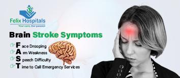
Recognizing the symptoms of a brain stroke is critical for getting prompt medical attention. The acronym F.A.S.T. is often used to remember the most common Brain stroke symptoms.

A concise guide explaining brain tumor surgery cost in Noida, including key factors, treatment options, and expert care.

Human metapneumovirus (HMPV) is a type of common respiratory virus that mimics the symptoms of the common cold. It belongs to the Paramyxoviridae family, closely related to the viruses that cause measles and mumps. While most cases infected by the virus are mild, some people are likely to get severely sick, especially the first time they get HMPV infection.
The HMPV infection is more common in winters and early spring but can occur at any time of the year. It affects people of all age groups, although infants and young children, older adults, and people with weakened immune systems are more vulnerable. Untreated cases of HMPV can develop into serious complications, such as pneumonia or bronchitis, therefore if your symptoms last for more than two weeks or become severe, seek medical help immediately.
Whether you’re looking to safeguard your family against the virus or seeking treatment for your child’s existing pediatric human metapneumovirus infection, understanding HMPV is the first step towards effective care.
For HMPV diagnosis, treatment or care, consult Felix Hospital, the best hospital for human metapneumovirus treatment in Noida and Greater Noida.
Human Metapneumovirus is highly contagious and spreads through respiratory droplets, close contact, or contaminated surfaces. Also, once HMPV enters your body, the incubation period lasts between three to six days.
Impact on Adults: Human Metapneumovirus infections in adults can range from mild cold-like symptoms to severe respiratory distress in high-risk groups, such as those with chronic lung diseases and cancer patients.
Impact on Children: Pediatric human metapneumovirus infection is particularly concerning, as it can cause bronchiolitis and pneumonia in infants and toddlers.
Like any virus, human metapneumovirus is contagious, which means it gets transmitted from an infected person. You might get exposed to the virus if you:
Touch surfaces that contain the virus, such as door handles, phones, etc.
Touch your mouth, nose, or eyes after you touch infected surfaces
Come in contact with respiratory droplets from an infected person. It could be through sneezing, spitting, or coughing
Come in direct contact with infected person, such as shaking hands, hugging and touching
Human metapneumovirus is seasonal and is more prevalent during winter or early spring months, same time as flu season.
Infants, young children, and older adults are at greater risk due to weaker immune defenses.
Individuals with pre-existing conditions such as asthma, chronic obstructive pulmonary disease (COPD), or other respiratory conditions are at heightened risk.
Individuals with weakened immune systems, such as those undergoing chemotherapy, are more vulnerable.
The symptoms of HMPV often mimic those of the flu or common cold that usually go away between two to five days. The symptoms can vary in severity depending on the individual’s age, immune status, and overall health.
Cough
Sore throat
Fever
Wheezing
Headache
If left untreated, symptoms can worsen and cause serious health issues in more severe cases, or patients with compromised immune systems. They may experience
Difficulty breathing
Asthma flare-ups
Respiratory infections such as bronchitis or pneumonia
Unless you’re very sick and hospitalized with HMPV, there’s a high chance that you won’t even know that you've been infected with the virus. However, diagnosing HMPV involves a combination of clinical evaluation and diagnostic testing:
Physical Examination: Your doctor will look for symptoms like wheezing, fever, and nasal discharge.
Swab Test: A soft-tipped stick (swab) is used to get a sample from your nose or throat. The sample is sent to the lab for confirmation of the virus.
Blood Tests: Complete blood counts (CBC) and inflammatory markers help evaluate the severity of the infection.
Molecular Tests: Reverse transcription-polymerase chain reaction (RT-PCR) tests detect the presence of HMPV RNA in respiratory samples.
Antigen Detection: Rapid antigen tests help identify the virus, especially in acute cases.
Bronchoscopy/Chest X-rays: Imaging may be required to rule out pneumonia or other complications in the airways of your lungs.
Currently, there’s no treatment for an upper respiratory infection caused by HMPV. Most people can easily manage their symptoms at home and rest until they feel better. The treatment for HMPV primarily focuses on management of symptoms that includes:
Coping with Symptoms: Take over-the-counter medications and cough syrups for fever, cough and pain. Nasal decongestants and saline nasal sprays can be used for congestion in the nose.
Hydration and Rest: Make sure you drink plenty of water and there's adequate fluid intake. Rest is crucial for recovery.
Oxygen Therapy: For severe cases involving respiratory distress, supplemental oxygen may be required.
Hospitalization: Pediatric or elderly patients with complications may need hospitalization.
At Felix Hospitals, the best hospital for human metapneumovirus treatment in Noida, our specialists provide comprehensive care, ensuring patients receive the tailored treatments and care.
You can reduce your risk of getting HMPV by taking these preventive measures:
Washing/Sanitizing your hands often with soap and water or hand sanitizer.
Cover your nose and mouth with your elbow while sneezing or coughing.
Avoid being around or coming in physical contact with an infected person.
Wearing a mask if you’re sick.
Regularly clean and disinfect high-touch surfaces like doorknobs and mobile phones.
Consume a balanced diet, exercise regularly, and avoid smoking to strengthen your immune system.
Don’t share towels, food or eating utensils with others.
Mild cases of human metapneumovirus usually last 3 to 7 days. If you’re very sick, it’ll probably take longer to recover. You might also have lingering symptoms, like a cough, that may take a little longer to go away.
At Felix Hospitals, we have the best doctors for human metapneumovirus (HMPV) treatment in Noida. Our multidisciplinary team combines expertise in pulmonology, internal medicine, pediatrics, and infectious diseases to deliver exceptional care. Leading the way, Dr. Priyadarshi Jitendra Kumar works alongside our highly skilled internal medicine specialists, including Dr. Anshumala Sinha, Dr. Sonakshi Saxena, Dr. Apoorva Shetty, Dr. Neelabh Pratap, Dr. Priyanka Singh, Dr. Ravi Sharma, and Dr. Keshav Kumar Garg.
Whether you need an outpatient consultation or inpatient care, our state-of-the-art facilities and personalized treatment plans ensure a seamless recovery process. At Felix Hospitals, your health is our priority.
Protect yourself and your loved ones from human metapneumovirus. Visit Felix Hospitals, the best hospital for human metapneumovirus treatment, and take proactive steps to safeguard your respiratory health.
Human metapneumovirus is a virus that causes upper and lower respiratory infections. While there’s no cure or vaccine for human metapneumovirus, most cases recover within a few days. However, sometimes infants, older people, and people who have weakenes immune systems can develop complications, like pneumonia. Therefore, understanding its symptoms, diagnosis, and treatment options is vital for effective management of HMPV. By taking preventive measures and seeking timely medical care, you can reduce the risk of complications and ensure a faster recovery.
Q1- How long does it take to recover from human metapneumovirus?
ANS: It largely depends on the individual’s overall health, age, and immunity. It’s crucial for anyone with existing respiratory symptoms, or compromised immune system to seek medical advice.
Q2- When should I see my doctor for pediatric human metapneumovirus infection?
ANS: You should seek medical advice if your child’s symptoms don’t start to improve within a few days or if you or your child has a fever lasting longer than three days. If your child has symptoms of a respiratory infection and an underlying condition that puts them at an elevated risk for severe illness, do not delay seeking treatment for pediatric human metapneumovirus infection.
Q3- Do I need antibiotics for human metapneumovirus?
ANS: No, HMPV is a virus and antibiotics only treat bacteria. However, if your doctor prescribes antibiotics, it would be to treat any secondary infections.
4- What are the complications of human metapneumovirus infections in adults?
ANS: Sometimes HMPV causes complications such as asthma, COPD flare-ups, pneumonia and bronchitis.

Felix Hospital in Noida is well known for its general practitioners who offer holistic treatment for different health issues, like the common cold and coughs.

Felix Hospital in Noida is esteemed as a leading institution for general medicine, particularly renowned for its excellence in treating cold and cough
For those experiencing such symptoms, seeking care from the best hospital for pulmonology treatment in Noida ensures you receive timely and expert diagnosis and treatment.

Dr. Priyadarshi Jitender Kumar stands out as a leading expert in diagnosing and treating pulmonary embolism.
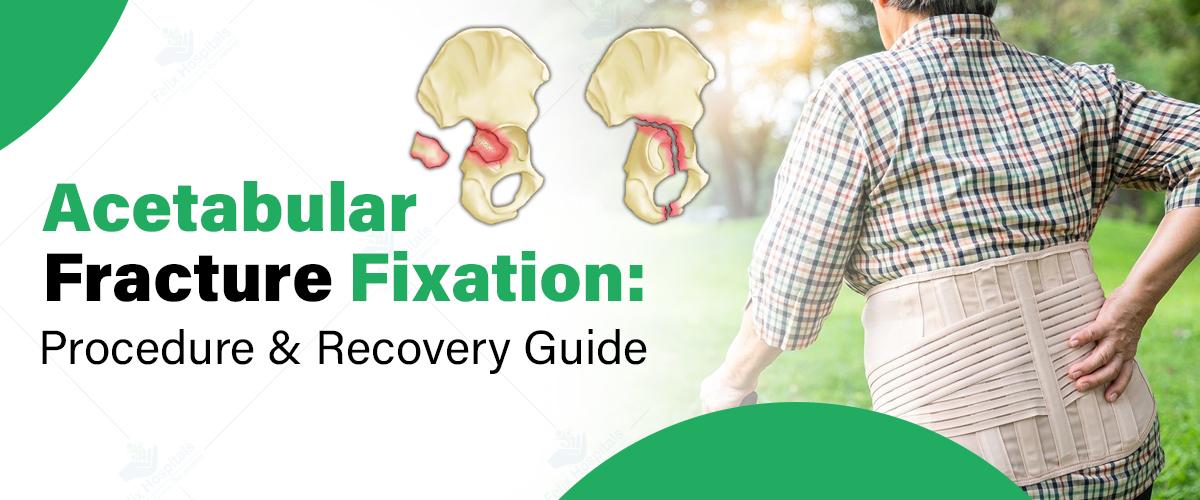
Acetabular fractures involve damage to the socket portion of the hip joint (the acetabulum), which plays a crucial role in maintaining the stability of the hip. This type of fracture is often the result of high-impact trauma, such as car accidents or falls from significant heights. Given the complexity of these fractures, proper fixation is vital for long-term recovery and to prevent complications that could affect mobility and quality of life. Seeking treatment at the best orthopedic hospital in Noida can significantly improve outcomes, as specialized care, advanced imaging, and experienced surgeons are crucial in managing such injuries. Understanding the procedure for acetabular fracture fixation and the recovery timeline is essential for patients to achieve the best possible results and regain their mobility.
Start Your Recovery Journey Now! Contact us at +91 9667064100 with our specialists in Noida and Greater Noida.
An acetabular fracture refers to a break in the acetabulum, which is the cup-like structure in the pelvis that forms the socket of the hip joint. These fractures are typically caused by significant trauma, such as motor vehicle accidents, falls, or sports-related injuries. The acetabulum works in conjunction with the femoral head (the ball portion of the hip joint) to provide a stable and functional hip joint.
There are several types of acetabular fractures, including:
Certain factors increase the risk of acetabular fractures, including:
Surgical fixation is typically necessary when the fracture is displaced or the hip joint is unstable. In cases where the fracture fragments misalign and cannot heal properly on their own, surgery is crucial. Several factors influence the decision to perform surgery, including:
Early intervention plays a key role in the recovery process. Fixing the fracture promptly improves the chances of a full recovery, reduces the risk of complications, and enhances long-term mobility.
Before surgery, the patient undergoes several diagnostic tests, including X-rays, CT scans, and physical exams. These tests help the surgeon assess the severity of the fracture and plan the best approach for fixation.
There are two primary types of fixation methods:
Surgical anesthesia options include general anesthesia, where the patient is fully asleep, or regional anesthesia, where only the lower body is numbed.
Immediately after surgery, the patient is monitored in a recovery room for signs of complications. Pain management is essential in this phase, and the medical team administers medication to keep discomfort under control. The hospital stay typically lasts several days, depending on the complexity of the surgery and the patient’s overall health.
Physical therapy begins as soon as possible to improve joint mobility, muscle strength, and overall functionality. The rehabilitation process is crucial to restoring the hip's full range of motion and preventing long-term complications. Patients are also monitored for potential complications, such as infections, blood clots, or issues with the fixation hardware (e.g., screws or plates).
The recovery timeline for acetabular fracture fixation varies based on the severity of the fracture, the patient’s age, and other health factors. It generally unfolds in three stages:
1. Immediate Post-Surgery Phase (First Few Days to Weeks): During this phase, patients are encouraged to rest and follow instructions regarding weight-bearing restrictions. Crutches or a walker may be required to avoid placing weight on the hip joint. Swelling, bruising, and discomfort are common, and patients typically remain in the hospital for a few days.
2. Short-Term Recovery (6 Weeks to 3 Months): At this stage, physical therapy plays an important role in regaining mobility. Patients gradually begin to increase weight-bearing on the affected hip, with the goal of achieving some level of mobility. X-rays are often performed to ensure the fracture is healing correctly. This phase may involve limited weight-bearing and assisted walking.
3. Long-Term Recovery (6 Months to 1 Year): Complete recovery from an acetabular fracture can take up to one year. During this time, patients work on strengthening the muscles surrounding the hip and improving flexibility. Full weight-bearing activities and a return to normal activity levels can occur, depending on the success of the surgery and the patient’s adherence to rehabilitation protocols.
Physical therapy is essential throughout the recovery process. Early exercises focus on improving flexibility and preventing joint stiffness, while later exercises emphasize building strength in the hip and leg muscles. The rehabilitation team will tailor exercises to the patient's progress and limitations. Full restoration of mobility is typically achieved within 6 to 12 months, but each patient’s timeline may vary.
During recovery, lifestyle adjustments may be necessary, such as avoiding high-impact activities (e.g., running, jumping) that could stress the healing joint. Nutritional considerations are also crucial; calcium and vitamin D-rich foods can promote bone healing. Maintaining bone health through weight-bearing exercises (when appropriate) and fall prevention strategies is key in avoiding future fractures.
Felix Hospitals is proud to offer expert care for acetabular fracture fixation, ensuring the best possible outcomes for patients. Whether you're seeking treatment in Noida or Greater Noida, our team of skilled orthopedic surgeons is equipped to provide personalized care tailored to your needs.
At Felix Hospital in Sector 137, Noida, patients can trust the expertise of our renowned orthopedic specialists:
These professionals bring extensive experience and advanced techniques to ensure a smooth recovery and excellent results.
For those in Greater Noida, Dr. Varun Aggrawal serves patients at Felix Hospital Gamma-1, Greater Noida. Known for his commitment to excellence, Dr. Aggrawal provides world-class orthopedic care to address complex acetabular fractures effectively.
Book an Appointment Today with the best orthopedic surgeons at Felix Hospitals today for expert care and personalized treatment.
Acetabular fracture fixation is a complex procedure, but when performed at a skilled orthopedic center, it offers patients a chance at a full recovery. A well-structured recovery plan that includes surgical intervention, post-operative care, and rehabilitation is crucial for regaining mobility and function. Adhering to medical advice and taking proactive steps during recovery can help patients return to their normal lives and reduce the risk of future complications. Seeking care at the best hospital for Acetabular fracture fixation in Noida ensures that patients receive the expertise and support they need for optimal healing.
1. What is an acetabular fracture?
Ans: An acetabular fracture is a break in the cup-like structure of the pelvis that forms the socket of the hip joint. This type of fracture typically results from high-impact trauma such as car accidents or falls.
2. When is surgery required for an acetabular fracture?
Ans: Surgical fixation is necessary when the fracture is displaced, the hip joint is unstable, or the bones cannot heal properly on their own. Surgery helps restore alignment and ensures proper healing.
3. What are the risks of acetabular fracture surgery?
Ans: While rare, potential risks include infection, blood clots, nerve or blood vessel damage, and issues with the fixation hardware. Choosing an experienced orthopedic surgeon can minimize these risks.
4. How long does it take to recover from acetabular fracture fixation surgery?
Ans: Recovery can take 6 to 12 months, depending on the severity of the fracture, the patient’s age, and adherence to rehabilitation protocols. Physical therapy is crucial for regaining strength and mobility.
5. What should I avoid during recovery from acetabular fracture fixation?
Ans: Avoid high-impact activities, such as running or jumping, until fully healed. Follow your doctor’s instructions regarding weight-bearing restrictions and maintain a diet rich in calcium and vitamin D for bone healing.
6. Can an acetabular fracture heal without surgery?
Ans: In cases where the fracture is non-displaced and the hip joint remains stable, non-surgical treatment may be an option. However, most acetabular fractures require surgical intervention for optimal healing.
7. Why should I choose Felix Hospitals for acetabular fracture fixation?
Ans: Felix Hospitals offers advanced imaging, skilled orthopedic surgeons, and personalized care to ensure the best outcomes for acetabular fractures.

Bone fractures are common injuries from accidents or falls. While many heal well, complications can arise, affecting recovery. Learn more about these issues and treatments.

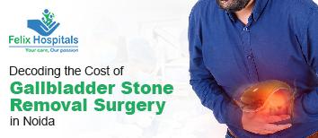
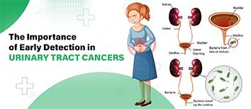
गॉल ब्लैडर (पित्ताशय) में पथरी या स्टोन होना महिलाओं में एक सामान्य समस्या बनती जा रही है। गॉल ब्लैडर, जो पित्त (बाइल) को संग्रहित और नियंत्रित करता है, पाचन में सहायक होता है। लेकिन कई बार यहां कोलेस्ट्रॉल या बिलीरुबिन के क्रिस्टल जमने लगते हैं, जिससे स्टोन बनने की संभावना बढ़ जाती है इसलिए समय रहते आप अच्छे ब्लैडर स्टोन ट्रीटमेंट हॉस्पिटल से समपर्क करें। इस ब्लॉग में हम गॉल ब्लैडर स्टोन के लक्षण, कारण और प्रभावी उपचार के बारे में जानेंगे, ताकि महिलाएं इस समस्या को समय रहते पहचान सकें और इलाज करा सकें।
ज्यादा जानकारी के लिए हमें कॉल करें +91 9667064100.
गॉल ब्लैडर स्टोन (पित्ताशय की पथरी) महिलाओं में एक सामान्य समस्या है, जिसमें गॉल ब्लैडर में पत्थर जैसी संरचनाएं बनने लगती हैं। गॉल ब्लैडर यकृत के नीचे स्थित एक छोटा अंग है, जो पित्त को संग्रहित करता है। यह पित्त वसा के पाचन में मदद करता है। जब बाइल में कोलेस्ट्रॉल, बिलीरुबिन, या अन्य पदार्थों की अधिकता हो जाती है, तो ये पदार्थ ठोस हो कर छोटे पत्थर जैसे रूप में बदल जाते हैं, जिन्हें पित्ताशय की पथरी कहते हैं। गॉल ब्लैडर स्टोन का समय पर उपचार करवाना बहुत जरूरी होता है, अन्यथा यह संक्रमण और गंभीर स्वास्थ्य समस्याओं का कारण बन सकता है।
महिलाओं में गॉल ब्लैडर स्टोन के लक्षण कई प्रकार के हो सकते हैं और अक्सर दर्द या अन्य असुविधाजनक स्थितियों के रूप में सामने आते हैं। यहां कुछ सामान्य लक्षण दिए गए हैं:
यह सबसे आम लक्षण है, जिसे बाइलिक कोलिक कहते हैं। दर्द अचानक आता है और हल्के से लेकर तीव्र तक हो सकता है, जो कुछ मिनटों से लेकर कई घंटों तक रह सकता है।
यह दर्द अक्सर दाएं कंधे और पीठ के दाईं ओर फैल सकता है।
कुछ महिलाओं को भोजन के बाद विशेष रूप से वसा युक्त भोजन के बाद मतली और उल्टी की समस्या हो सकती है।
पथरी से प्रभावित महिलाओं को वसा युक्त भोजन के बाद पेट में भारीपन, सूजन, और गैस की समस्या हो सकती है।
यदि पथरी के कारण संक्रमण हो गया है, तो तेज बुखार, पसीना, और ठंड लगने की समस्या भी हो सकती है।
यदि पथरी बाइल डक्ट (पित्त नलिका) को अवरुद्ध कर देती है, तो त्वचा और आंखों का रंग पीला हो सकता है।
अवरुद्ध पित्त नलिका के कारण, मल का रंग हल्का और यूरिन का रंग गहरा हो सकता है।
गॉल ब्लैडर स्टोन के लक्षणों का अनुभव हर महिला में अलग हो सकता है और गंभीरता में भिन्न हो सकते हैं। अगर लक्षण लगातार और तीव्र हो रहे हों, तो तुरंत चिकित्सा परामर्श लेना जरूरी होता है, क्योंकि इससे संक्रमण या अन्य जटिलताएं उत्पन्न हो सकती हैं।
महिलाओं में गॉल ब्लैडर स्टोन मुख्य रूप से दो प्रकार के होते हैं, जो पत्थर की संरचना और उनकी उत्पत्ति के आधार पर अलग-अलग होते हैं:
ये गॉल ब्लैडर स्टोन का सबसे सामान्य प्रकार हैं, और ज्यादातर महिलाओं में पाए जाते हैं। कोलेस्ट्रॉल स्टोन पीले या हरे रंग के होते हैं। ये तब बनते हैं जब बाइल में कोलेस्ट्रॉल की अधिकता होती है, जो घुल नहीं पाता और ठोस कणों में बदल जाता है। इसका कारण खराब आहार, मोटापा और हार्मोनल बदलाव हो सकता है, जैसे गर्भावस्था के दौरान।
ये गहरे भूरे या काले रंग के होते हैं और तब बनते हैं जब बाइल में बिलीरुबिन की मात्रा बढ़ जाती है। बिलीरुबिन एक पिगमेंट है जो शरीर में लाल रक्त कोशिकाओं के टूटने से बनता है। पिगमेंट स्टोन अक्सर उन महिलाओं में बनते हैं जो लिवर की बीमारियों जैसे सिरोसिस, रक्त विकार जैसे सिकल सेल एनीमिया, या बाइल डक्ट में संक्रमण से पीड़ित होती हैं।
महिलाओं में गॉल ब्लैडर स्टोन के कई कारण हो सकते हैं, जिनमें हार्मोनल बदलाव, खान-पान की आदतें, और जीवनशैली से जुड़े कई कारक शामिल हैं। यहां कुछ प्रमुख कारण दिए गए हैं:
इन कारणों को ध्यान में रखकर यदि महिलाएं अपने खान-पान और जीवनशैली में बदलाव करती हैं, तो गॉल ब्लैडर स्टोन की संभावना को कम किया जा सकता है।
गॉल ब्लैडर स्टोन से बचाव और निदान के लिए कुछ एहतियात और जांच प्रक्रियाएं अपनाई जा सकती हैं। समय पर पहचान और सही उपचार से इस समस्या को रोका और नियंत्रित किया जा सकता है।
वसा और कोलेस्ट्रॉल की मात्रा कम रखें। अधिक तले हुए, वसायुक्त और जंक फूड से बचें। फाइबर युक्त खाद्य पदार्थ जैसे सब्जियां, फल और साबुत अनाज का सेवन करें। चीनी और रिफाइंड कार्बोहाइड्रेट का सेवन सीमित करें।
अधिक वजन गॉल ब्लैडर स्टोन का खतरा बढ़ा सकता है। स्वस्थ तरीके से वजन कम करें और नियमित व्यायाम करें।
नियमित शारीरिक गतिविधि बाइल का प्रवाह बनाए रखती है, जिससे पथरी बनने का खतरा कम होता है।
अनियमित भोजन और अधिक लंबे समय तक भूखे रहना गॉल ब्लैडर स्टोन की संभावना बढ़ा सकता है। सही समय पर भोजन करें।
ये आदतें यकृत और गॉल ब्लैडर की कार्यक्षमता को प्रभावित कर सकती हैं और पथरी की संभावना को बढ़ा सकती हैं।
अगर आप हार्मोनल दवाएं ले रही हैं या गर्भनिरोधक गोलियों का उपयोग कर रही हैं, तो डॉक्टर से परामर्श लें, क्योंकि ये पथरी के खतरे को बढ़ा सकती हैं।
यदि लक्षण महसूस होते हैं, तो डॉक्टर कुछ जांचों की सलाह दे सकते हैं ताकि गॉल ब्लैडर स्टोन का पता लगाया जा सके:
यह सबसे सामान्य और प्रभावी जांच है जो गॉल ब्लैडर में स्टोन की उपस्थिति की पुष्टि करती है। अल्ट्रासाउंड से पथरी का आकार, संख्या, और स्थान ज्ञात होता है।
यह जांच गॉल ब्लैडर की कार्यक्षमता को मापती है और यह पता लगाने में मदद करती है कि बाइल ब्लैडर से सही तरीके से निकल रहा है या नहीं।
सीटी स्कैन से गॉल ब्लैडर और बाइल डक्ट की विस्तार से तस्वीरें ली जाती हैं। इससे छोटे स्टोन भी दिखाई दे सकते हैं।
यह एमआरआई की तरह एक विशेष स्कैन है, जो बाइल डक्ट्स और गॉल ब्लैडर की विस्तृत छवि दिखाता है और जटिल मामलों में उपयोगी होता है।
ब्लड टेस्ट:
ब्लड टेस्ट से संक्रमण, पीलिया, और बाइल डक्ट के अवरोध का पता लगाया जा सकता है। इसमें बिलीरुबिन, लिवर एंजाइम, और सफेद रक्त कोशिकाओं की मात्रा देखी जाती है।
अगर गॉल ब्लैडर स्टोन की पुष्टि हो जाती है और लक्षण गंभीर हैं, तो डॉक्टर सर्जरी की सलाह दे सकते हैं। हल्के लक्षणों वाले मामलों में, लाइफस्टाइल में सुधार और आहार पर ध्यान देकर पथरी को बढ़ने से रोका जा सकता है।
गॉल ब्लैडर स्टोन का जल्दी निदान और उचित बचाव पथरी के कारण होने वाले दर्द और अन्य जटिलताओं को रोक सकता है।
महिलाओं में गॉल ब्लैडर स्टोन का उपचार लक्षणों की गंभीरता, पथरी के आकार, और स्थिति पर निर्भर करता है। यहां कुछ प्रभावी उपचार विकल्प दिए गए हैं:
लेप्रोस्कोपिक कोलेसिस्टेक्टॉमी: यह एक न्यूनतम इनवेसिव सर्जरी है जिसमें पेट में छोटे-छोटे चीरे लगाए जाते हैं और गॉल ब्लैडर को निकाल दिया जाता है। यह सबसे आम और सुरक्षित प्रक्रिया है, जिसमें अस्पताल में कुछ ही दिन रुकना पड़ता है।
यदि लेप्रोस्कोपिक सर्जरी संभव नहीं होती या जटिलताएं होती हैं, तो ओपन सर्जरी का सहारा लिया जाता है। इसमें पेट में बड़ा चीरा लगाया जाता है, जिससे मरीज को ठीक होने में थोड़ा अधिक समय लगता है।
यदि सर्जरी संभव नहीं है, तो कुछ दवाएं (जैसे उर्सोडिऑल या केनोडिऑल) दी जाती हैं जो कोलेस्ट्रॉल स्टोन को घोलने में मदद कर सकती हैं। हालांकि, इस उपचार में समय लगता है और यह छोटे कोलेस्ट्रॉल स्टोन के लिए ही प्रभावी होता है। यह तरीका पिगमेंट स्टोन के लिए उतना कारगर नहीं होता।
अगर पथरी बाइल डक्ट में अटक जाती है और संक्रमण या जटिलताओं का कारण बनती है, तो ईआरसीपी की मदद से इसे हटाया जाता है। इस प्रक्रिया में एंडोस्कोप के माध्यम से बाइल डक्ट से पथरी को बाहर निकाला जाता है। यह एक सर्जरी नहीं है, लेकिन इसमें विशेषज्ञता की आवश्यकता होती है।
यह एक गैर-सर्जिकल प्रक्रिया है जिसमें उच्च-ऊर्जा वाली शॉक वेव्स का उपयोग करके गॉल ब्लैडर स्टोन को छोटे-छोटे टुकड़ों में तोड़ा जाता है, ताकि वे बाइल के साथ निकल सकें। यह प्रक्रिया बहुत आम नहीं है और इसे केवल छोटे कोलेस्ट्रॉल स्टोन के लिए ही उपयोग किया जाता है।
अगर पथरी छोटी है और गंभीर लक्षण नहीं है, तो आहार और जीवनशैली में सुधार से इसे नियंत्रित किया जा सकता है। वसा युक्त खाद्य पदार्थों से बचें, संतुलित आहार लें, और नियमित व्यायाम करें। इससे गॉल ब्लैडर के स्वास्थ्य में सुधार हो सकता है और पथरी के बढ़ने की संभावना कम होती है।
कुछ लोग सेब का रस, सेब का सिरका, या हल्दी का उपयोग करते हैं, जो बाइल फ्लो में सुधार ला सकते हैं। हालांकि, इनका वैज्ञानिक प्रमाण सीमित है, इसलिए किसी भी घरेलू उपचार को अपनाने से पहले डॉक्टर से परामर्श लेना चाहिए।
अगर गॉल ब्लैडर स्टोन का आकार बढ़ रहा है या लक्षण बिगड़ रहे हैं, तो नियमित जांच और डॉक्टर की सलाह पर सर्जरी या अन्य प्रक्रियाएं करवाना सही रहता है।
गॉल ब्लैडर स्टोन का प्रभावी उपचार का चयन स्थिति, लक्षण, और स्वास्थ्य पर निर्भर करता है। सही उपचार व नोएडा में इलाज की कीमत के बारे में जानने के लिए डॉक्टर से परामर्श लेना सबसे महत्वपूर्ण है, ताकि पथरी से होने वाली संभावित जटिलताओं से बचा जा सके।
गॉल ब्लैडर स्टोन के इलाज के लिए कुछ विशेष प्रकार के डॉक्टरों और विशेषज्ञों की आवश्यकता होती है, जो इस स्थिति की पहचान, निदान, और उपचार में मदद करते हैं।
डॉ. रितेश अग्रवाल: फेलिक्स हॉस्पिटल में लैप्रोस्कोपिक सर्जरी में विशेषज्ञता रखने वाले प्रमुख सर्जन हैं, जो मिनिमली इनवेसिव प्रक्रियाओं के जरिए जटिल सर्जिकल मामलों का इलाज करते हैं।
डॉक्टर की सलाह के लिए आज ही फोन करें +91 9667064100.
महिलाओं में गॉल ब्लैडर स्टोन की समस्या आम होती जा रही है, लेकिन इसे पहचानने और सही उपचार के जरिए इससे बचा जा सकता है। समय पर डॉक्टर से परामर्श लेकर सही उपचार करवाना बेहद जरूरी है, ताकि पथरी से होने वाली किसी भी जटिलता को रोका जा सके। स्वस्थ जीवनशैली और नियमित स्वास्थ्य जांच से आप इस समस्या से काफी हद तक बच सकती हैं। इस जानकारी से आपको गॉल ब्लैडर स्टोन के बारे में सही निर्णय लेने में मदद मिलेगी और इस समस्या से निपटने में सहायता मिलेगी।
प्रश्न 1: महिलाओं में गॉल ब्लैडर स्टोन क्यों होते हैं ?
उत्तर: हार्मोनल बदलाव, विशेषकर एस्ट्रोजन का बढ़ा हुआ स्तर, अनुवांशिकता, मोटापा, अधिक कोलेस्ट्रॉल वाला आहार, और तेजी से वजन घटाना महिलाओं में गॉल ब्लैडर स्टोन के सामान्य कारण हैं।
प्रश्न 2: गर्भावस्था में गॉल ब्लैडर स्टोन का खतरा बढ़ता है?
उत्तरः हां, गर्भावस्था में एस्ट्रोजन और प्रोजेस्टेरोन के स्तर में बदलाव होने से गॉल ब्लैडर में कोलेस्ट्रॉल की मात्रा बढ़ सकती है, जिससे स्टोन बनने की संभावना बढ़ जाती है।
प्रश्न 3: क्या गॉल ब्लैडर स्टोन का इलाज बिना सर्जरी के संभव है ?
उत्तर: छोटे कोलेस्ट्रॉल स्टोन के लिए दवाइयां और शॉक वेव लिथोट्रिप्सी जैसी विधियों से इलाज संभव है, लेकिन बड़े स्टोन और गंभीर लक्षणों के लिए सर्जरी की आवश्यकता हो सकती है।
प्रश्न 4: गॉल ब्लैडर स्टोन से कैसे बचा जा सकता है ?
उत्तर: वसा और कोलेस्ट्रॉल युक्त खाद्य पदार्थों का सेवन कम करें, संतुलित आहार लें, नियमित व्यायाम करें, और वजन को नियंत्रित रखें। यह सब गॉल ब्लैडर स्टोन से बचाव में सहायक हो सकता है।
प्रश्न 5: क्या गॉल ब्लैडर हटाने से कोई दुष्प्रभाव होता है ?
उत्तर: गॉल ब्लैडर हटाने के बाद शरीर में बाइल को सीधे छोटी आंत में छोड़ा जाता है, जिससे कुछ लोगों को पाचन में हल्की समस्याएं हो सकती हैं, लेकिन अधिकतर लोग सामान्य जीवन जी सकते हैं।
प्रश्न 6: गॉल ब्लैडर स्टोन की पहचान कैसे होती है ?
उत्तर: अल्ट्रासाउंड, सीटी स्कैन, या एमआरआई जैसी इमेजिंग तकनीक से गॉल ब्लैडर स्टोन की पहचान की जा सकती है। गंभीर मामलों में ईआरसीपी भी की जाती है।
प्रश्न 7: क्या गॉल ब्लैडर स्टोन कैंसर का कारण बन सकता है ?
उत्तर: लंबे समय तक गॉल ब्लैडर में स्टोन रहने से गॉल ब्लैडर कैंसर का जोखिम बढ़ सकता है, लेकिन ऐसा बहुत दुर्लभ होता है। गॉल ब्लैडर स्टोन की सही समय पर पहचान और इलाज से इस खतरे को कम किया जा सकता है।
प्रश्न 8: क्या गॉल ब्लैडर स्टोन के साथ सामान्य जीवन संभव है ?
उत्तर: यदि स्टोन से कोई गंभीर लक्षण नहीं हैं, तो कुछ लोग गॉल ब्लैडर स्टोन के साथ भी सामान्य जीवन जी सकते हैं। लेकिन समय-समय पर डॉक्टर से जांच कराते रहना आवश्यक है ताकि जटिलताओं से बचा जा सके।

Gallbladder stone surgery, or cholecystectomy, is typically performed when gallstones lead to significant health issues.

When faced with a health issue, knowing where to seek care—whether at a polyclinic or a hospital—can be crucial for timely and effective treatment. The choice often depends on factors such as affordability, convenience, type of illness, and access to specialized medical knowledge. Let’s explore the differences between polyclinics and hospitals, helping you understand which option may be best suited for your health needs.
Trust Felix Hospitals for the best care, specialized medical services, and compassionate support during your critical moments by Clicking Here.
A polyclinic is a healthcare facility that offers a range of outpatient services, usually from general practitioners and specialists. It is typically smaller than a hospital but provides comprehensive care, including diagnostic tests, preventive care, and minor procedures. Polyclinics are usually situated in communities or near residential areas, making them accessible for routine consultations and minor health issues.
Services Provided: Routine check-ups, vaccinations, minor surgeries, lab tests, and general consultations.
Doctors and Specialists: Often includes general practitioners, dermatologists, dentists, and other specialists.
Affordability: Polyclinics tend to be more affordable than hospitals, making them a preferred choice for non-urgent care.
Hospitals are large medical facilities equipped to handle a wide range of health conditions, from routine check-ups to emergency care and complex surgeries. Hospitals often include multiple departments with advanced diagnostic and treatment facilities, inpatient care, and specialized intensive care units (ICUs).
Services Provided: Emergency care, surgeries, intensive care, specialized treatments, and inpatient care.
Doctors and Specialists: Hospitals employ a wider range of healthcare professionals, including surgeons, anesthesiologists, cardiologists, and more.
Affordability: Due to their advanced facilities and range of services, hospitals are generally more expensive than polyclinics.
Polyclinic: Polyclinics are usually more cost-effective for patients needing general consultations, minor treatments, or routine screenings. They are ideal for individuals looking to manage healthcare costs for non-emergency issues.
Hospital: Hospitals tend to be more expensive due to the availability of specialized care, emergency services, and inpatient facilities. Insurance coverage may reduce costs for certain procedures, but overall, hospitals are a more costly option.
Polyclinic: Located in or near residential areas, polyclinics are designed to be easily accessible. For minor issues or follow-up consultations, polyclinics can save time and effort, especially for routine visits.
Hospital: Hospitals are often located in central urban areas and may be further from residential neighborhoods. While they offer comprehensive care, traveling to a hospital may be less convenient for quick consultations or minor issues.
Polyclinic: For minor ailments like the common cold, seasonal flu, allergies, and minor injuries, a polyclinic is often sufficient. Polyclinics are also ideal for chronic disease management, preventive care, and follow-ups.
Hospital: For severe conditions, such as trauma, heart attacks, complex surgeries, or diseases requiring intensive care, the best hospitals in Noida are the better choice. Hospitals have the necessary equipment, specialists, and facilities for managing life-threatening conditions.
Polyclinic: Polyclinics usually have general practitioners and specialists for common issues. However, the depth of expertise may be limited compared to hospitals, which have specialists in nearly every area of medicine.
Hospital: Hospitals are more equipped with a broader array of specialists and advanced diagnostic technology, providing higher expertise for complex medical conditions. In a hospital setting, multidisciplinary teams often work together for more comprehensive care.
Polyclinic: Polyclinics provide a sense of continuity and familiarity, as patients often see the same doctors and staff for routine care, making it easier to track health history.
Hospital: While hospitals also offer follow-up care, they are better suited for episodic or specialized care. Patients may find it harder to receive continuity in terms of seeing the same physician repeatedly.
Not every health issue requires a trip to the hospital. For common illnesses, polyclinics are often sufficient and more affordable. Regular check-ups, treatment for colds, minor infections, skin issues, and chronic disease management are generally manageable in a polyclinic. Hospitals are ideal for conditions that are severe, unexpected, or require immediate attention and complex treatments.
Consider these factors when deciding where to seek care:
1. Severity of Symptoms: For severe or sudden symptoms (e.g., chest pain, high fever, severe injuries), go to a hospital. For minor symptoms, consider visiting a polyclinic.
2. Type of Condition: If you have a common or minor health condition, a polyclinic is more affordable and convenient. For complex or rare conditions, a hospital provides more specialized expertise.
3. Location and Travel Time: For frequent visits, choose a nearby polyclinic. For major procedures, the travel may be worthwhile if the hospital has specialized care options.
4. Budget and Insurance: Evaluate your budget and insurance coverage. Hospitals may be more costly but are often covered by insurance plans for major treatments.
5. Availability of Services: Some medical needs (e.g., surgeries) can only be performed in hospitals, whereas others, like general consultations, can be handled in polyclinics.
For routine health check-ups and vaccinations.
To manage chronic conditions like hypertension or diabetes.
For minor illnesses or injuries that don’t require hospitalization.
For diagnostic tests that do not need hospitalization.
Felix Hospitals operates three convenient polyclinics in Noida and Delhi, at Noida Sector 75, Noida Sector 135, and New Ashok Nagar in Delhi. These centers offer comprehensive healthcare services for all your routine and non-emergency medical needs, ensuring you receive the best care close to home.
For severe illnesses, such as heart attacks, major surgeries, or traumatic injuries.
For illnesses that require complex diagnosis, specialist care, or advanced equipment.
In situations that may need emergency treatment or intensive care.
For prolonged treatment that requires an inpatient stay.
Trust Felix Hospitals for the best care, specialized medical services, and compassionate support during your critical moments by Clicking Here.
Choosing between a polyclinic and a hospital can be a straightforward decision when you understand your health needs and the services each facility offers. Polyclinics are an excellent choice for everyday health needs, preventive care, and minor treatments, providing affordable and accessible healthcare options. Hospitals, on the other hand, are essential for emergencies, complex treatments, and specialized care. By understanding your medical condition, budget, and the type of care needed, you can make an informed decision and ensure you receive the right care at the right time.
1. What is the difference between a polyclinic and a hospital?
Ans: A polyclinic is typically an outpatient facility offering routine care, while a hospital offers a wide range of services, including emergency and inpatient care.
2. Are polyclinics cheaper than hospitals?
Ans: Yes, polyclinics are generally more affordable and ideal for minor or routine healthcare needs.
3. Can polyclinics handle emergencies?
Ans: Polyclinics may provide basic first aid, but severe emergencies are better handled in hospitals equipped with emergency departments.
4. Should I visit a hospital for a cold or fever?
Ans: For minor illnesses like a cold or fever, visiting a polyclinic is usually sufficient.
5. Which is better for chronic illness management?
Ans: Polyclinics are often suitable for regular monitoring of chronic conditions, though hospitals may offer specialized treatment options if needed.

Felix Healthcare Private Limiteds main goal is to provide top-notch healthcare services to individuals of their background.

In vitro fertilization (IVF) and intrauterine insemination (IUI) have revolutionized the field of reproductive medicine, offering hope to couples facing infertility challenges. Over the years, these techniques have evolved dramatically, driven by advancements in technology, research, and clinical practices. The innovations in IVF and IUI have improved success rates, reduced complications, and opened up new possibilities for those struggling to conceive. Let’s delve into the latest advancements in IVF and IUI technologies, exploring how these innovations have transformed fertility treatment with the help of the best gynecologist hospital in Noida.
Don't let pelvic pain disrupt your pregnancy journey. Schedule a consultation with our expert gynecologists today and receive the best possible care by Clicking Here.
Before diving into the innovations, it is essential to understand the basic principles of IVF and IUI. Both are assisted reproductive technologies (ART) designed to help individuals or couples conceive.
In recent years, significant advancements in both IVF and IUI have led to higher success rates, fewer side effects, and more personalized treatments. These innovations range from the development of better stimulation protocols to cutting-edge laboratory techniques that improve embryo selection and implantation outcomes.
One of the most groundbreaking advances in IVF is Preimplantation Genetic Testing (PGT). This technique allows embryos to be screened for genetic abnormalities before they are transferred into the uterus. By identifying chromosomal defects, PGT improves the chances of a healthy pregnancy and reduces the risk of miscarriage. It is especially beneficial for individuals with a family history of genetic disorders or those who have experienced recurrent pregnancy loss.
PGT is further classified into three types:
PGT-A (Aneuploidy Testing): Detects chromosomal abnormalities that may lead to miscarriage or genetic diseases.
PGT-M (Monogenic/Single Gene Testing): Screens for specific hereditary genetic conditions, such as cystic fibrosis or sickle cell anemia.
Traditionally, embryologists evaluated embryos at specific time intervals to determine their quality. However, time-lapse imaging has revolutionized the process by allowing continuous monitoring of embryos without disturbing them. This technology provides a detailed picture of embryo development, helping embryologists select the best embryos for transfer based on their growth patterns.
The introduction of time-lapse incubators has increased implantation rates and reduced the likelihood of multiple pregnancies, leading to more successful IVF outcomes.
Artificial Intelligence (AI) is another technological breakthrough in IVF. By analyzing vast amounts of data from previous IVF cycles, AI can predict which embryos are most likely to result in a successful pregnancy. AI-driven software can assess embryo quality more accurately than the human eye, making the selection process faster and more precise.
AI also plays a crucial role in optimizing ovarian stimulation protocols, ensuring the right dose and timing of medications for each patient, leading to better egg quality and retrieval rates.
The traditional approach in IVF was to transfer fresh embryos shortly after fertilization. However, research has shown that frozen embryo transfers (FET) often result in higher pregnancy rates compared to fresh transfers. The freeze-all strategy allows all embryos to be frozen and transferred in a subsequent cycle when the uterine environment is more favorable.
This approach has also reduced the risk of ovarian hyperstimulation syndrome (OHSS), a potentially dangerous complication of fertility treatments. By freezing all embryos and waiting for the ovaries to recover, patients can undergo the transfer in a more controlled and safer manner.
While conventional IVF involves high doses of hormonal medications to stimulate the ovaries, minimal stimulation IVF uses lower doses of drugs, resulting in fewer eggs being retrieved. Although fewer eggs are collected, the quality of the eggs tends to be higher, and the procedure is less physically demanding for the patient. This approach is gaining popularity, especially for women with poor ovarian reserve or those seeking a more natural treatment option.
The introduction of embryo glue has also contributed to higher implantation rates. This solution, rich in hyaluronan, is applied to the embryos before transfer to enhance their adhesion to the uterine lining. By mimicking the natural environment of the uterus, embryo glue improves the chances of successful implantation, particularly for women who have experienced previous failed IVF cycles.
Traditionally, IUI was performed without real-time imaging. However, ultrasound-guided IUI has become increasingly common, providing greater accuracy in sperm placement. By visualizing the uterus during the procedure, the doctor can ensure that the sperm is deposited closer to the fallopian tubes, increasing the chances of fertilization.
Ultrasound-guided IUI is particularly useful for women with anatomical issues or those undergoing multiple IUI cycles without success.
The success of IUI largely depends on the quality of the sperm used in the procedure. In recent years, improved sperm preparation techniques, such as density gradient centrifugation and swim-up method, have been developed. These methods isolate the healthiest and most motile sperm, increasing the chances of fertilization.
Additionally, sperm DNA fragmentation testing is now being used to assess sperm quality more comprehensively. This test measures the level of DNA damage in sperm cells, providing insight into whether the sperm can successfully fertilize an egg and lead to a viable pregnancy.
A more advanced version of IUI, intrauterine sperm injection (IUSI), involves injecting sperm directly into the fallopian tubes instead of the uterus. This technique increases the proximity of the sperm to the egg, making fertilization more likely. IUSI is particularly beneficial for couples with severe male infertility or unexplained infertility.
Advances in ovarian stimulation protocols have improved the success rates of IUI. New drugs and tailored protocols are now available to stimulate ovulation more effectively, increasing the number of eggs released during an IUI cycle. By closely monitoring hormone levels and adjusting medications, doctors can optimize the timing of insemination for better results.
As research continues to progress, the future of IVF and IUI holds even more promise. Some of the emerging technologies and concepts that are expected to shape the future of fertility treatments include:
1. Gene Editing
The advent of gene-editing technologies like CRISPR has opened up the possibility of correcting genetic mutations in embryos. While still in its early stages, gene editing could one day be used to eliminate inherited diseases from embryos, providing couples with the opportunity to have healthy children free from genetic disorders.
2. Stem Cell Therapy
Stem cell research is another exciting area with potential applications in fertility treatment. Scientists are exploring the possibility of using stem cells to create new eggs or sperm for individuals with low fertility. This could offer a new solution for women with diminished ovarian reserve or men with severe sperm defects.
3. Organoid Technology
In the future, organoid technology could be used to create artificial reproductive organs, such as ovaries or testes, to support fertility in individuals with damaged or non-functional reproductive systems. This would provide new hope for individuals who currently have no viable options for biological parenthood.
4. Improved Non-Invasive Testing
New non-invasive techniques for assessing embryo quality, such as analyzing embryo culture media, are being developed. These methods could provide valuable information about embryo
Felix Hospital, the best IUI center in Noida is here to help you. Our best Doctor for IVF and IUI treatment—Dr. Sangeeta Sharma, Dr. Charu Yadav, and Dr. Sonia Kuruvilla—provide compassionate, personalized care tailored to your specific needs. As leading gynecologists in Noida, we focus on your health, comfort, and overall well-being. Reach out to us today to schedule a consultation and get the specialized care you deserve throughout your pregnancy.
Experience compassionate, personalized pregnancy care. Contact us now to begin your journey toward a healthy, comfortable pregnancy by calling +91 9667064100.
The field of assisted reproductive technologies, particularly IVF and IUI, has made remarkable strides in recent years. From preimplantation genetic testing and time-lapse imaging to AI-driven embryo selection and advanced sperm preparation techniques, these innovations have significantly improved the success rates and safety of fertility treatments. As these technologies evolve, couples are also seeking information on the IVF and IUI cost in Noida, which varies depending on the clinic and personalized treatment protocols, making it important to understand all aspects of the process.
As technology continues to evolve, the future of IVF and IUI looks brighter than ever. With the ongoing development of gene editing, stem cell therapy, and non-invasive testing, the dream of parenthood is becoming more achievable for individuals and couples facing infertility challenges.
1. What causes pelvic pain during pregnancy?
Ans: Pelvic pain during pregnancy can be caused by several factors, including the growing uterus, hormonal changes, pressure on the pelvic floor muscles, and loosening joints. Conditions like pelvic girdle pain (PGP) or symphysis pubis dysfunction (SPD) may also contribute to discomfort.
2. Is pelvic pain during pregnancy normal?
Ans: While mild pelvic discomfort can be common due to the body’s changes during pregnancy, severe or persistent pelvic pain should be evaluated by a healthcare professional to rule out any serious conditions.
3. How can I manage pelvic pain at home?
Ans: You can manage pelvic pain at home by practicing gentle exercises, using supportive pelvic belts, applying heat packs, and maintaining good posture. However, always consult your doctor before starting any home treatments.
4. When should I see a doctor for pelvic pain?
Ans: You should consult a doctor if your pelvic pain is severe, sudden, or persistent, or if it's accompanied by symptoms like bleeding, fever, or difficulty walking. Prompt medical attention can help ensure the health and safety of both you and your baby.
5. Can pelvic pain affect labor or delivery?
Ans: Pelvic pain, particularly conditions like SPD, can impact labor or delivery, making it more uncomfortable or difficult. However, with proper care and medical management, most women can have a safe and healthy delivery.
6. What treatments are available for pelvic pain during pregnancy?
Ans: Treatment options may include physical therapy, pelvic support belts, pain relief medications (approved by your doctor), and lifestyle modifications. In more severe cases, a doctor may recommend specialized treatment plans tailored to your condition.
7. Can pelvic pain indicate preterm labor?
Ans: In some cases, pelvic pain may be a sign of preterm labor, especially if accompanied by symptoms like contractions, lower back pain, or vaginal discharge. If you suspect preterm labor, seek immediate medical attention.
8. Will pelvic pain continue after pregnancy?
Ans: In many cases, pelvic pain resolves after delivery as the body returns to its pre-pregnancy state. However, some women may experience lingering pain, and it’s essential to seek follow-up care if the discomfort persists postpartum.
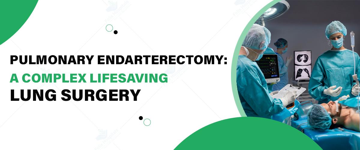
Pulmonary Endarterectomy (PEA) is a complex yet lifesaving surgical procedure for those suffering from chronic thromboembolic pulmonary hypertension (CTEPH). This surgery aims to remove blockages in the pulmonary arteries, improving blood flow and relieving symptoms such as shortness of breath and chest pain. For anyone considering this surgery, it's essential to choose the best hospital for Pulmonary Endarterectomy to ensure optimal outcomes and advanced post-operative care. Felix Hospital provides high-quality care with a team of experienced specialists, including Dr. Priyadarshi Jitendra Kumar, renowned for his expertise in pulmonology.
If you or a loved one is dealing with CTEPH, don’t hesitate to Click Here to reach out to Felix Hospital.
Pulmonary Endarterectomy (PEA) is a highly specialized surgical procedure used to treat CTEPH by removing organized blood clots from the pulmonary arteries. When blood clots from deep veins lodge in the pulmonary arteries, they obstruct blood flow and can lead to increased pressure in the lungs, resulting in CTEPH. PEA surgery removes these obstructions, allowing blood to flow freely, reducing lung pressure, and improving oxygenation.
Surgery is typically required in patients with CTEPH when:
1. Significant Blockage: Persistent blood clots obstruct the pulmonary arteries, causing increased pressure and symptoms.
2. Severe Symptoms: Patients experience severe shortness of breath, fatigue, or chest pain that impacts their quality of life.
3. Ineffectiveness of Medication: For some patients, medications are ineffective in managing CTEPH, making surgery the best option.
4. Risk of Heart Failure: Elevated lung pressure can lead to heart strain or right heart failure if untreated.
PEA is often a last-resort option, usually considered after confirming that other treatments or medications won’t suffice.
CTEPH primarily arises from unresolved pulmonary embolisms (PEs) or blood clots that form in the deep veins, usually in the legs, and travel to the lungs. When these clots do not dissolve, they can permanently lodge in the pulmonary arteries, causing long-term pressure and damage.
Other factors that may contribute to the development of CTEPH include:
Genetic predispositions
Chronic inflammatory or autoimmune conditions
Certain blood disorders
Previous surgeries or trauma
To accurately diagnose CTEPH and determine if PEA surgery is appropriate, a series of tests and imaging studies are performed:
1. Pulmonary Angiography: This imaging test provides detailed visuals of the pulmonary arteries to locate any obstructions.
2. Echocardiography: This test measures pressure within the pulmonary arteries and assesses heart function.
3. Ventilation-Perfusion (V/Q) Scan: A V/Q scan identifies regions in the lungs where blood flow may be restricted.
4. CT Pulmonary Angiography: CT angiography allows doctors to view blood flow within the pulmonary arteries and identify any blood clots.
5. Right Heart Catheterization: This procedure helps measure blood pressure in the lungs to confirm the presence of CTEPH.
The prognosis for patients undergoing PEA surgery is generally favorable, especially when treated at a well-equipped facility with expert surgeons. PEA provides substantial symptom relief, improved quality of life, and increased survival rates, though results can vary based on individual health factors. The cost of a Pulmonary Endarterectomy surgery depends on factors such as hospital facilities, surgeon fees, and post-operative care requirements. Patients are encouraged to discuss the expected costs with their healthcare provider to prepare for both the surgery and the recovery phase.
At Felix Hospital, we’re proud to have Dr. Priyadarshi Jitendra Kumar, one of the best pulmonologists, specializing in pulmonary surgeries and advanced respiratory care. Dr. Kumar’s extensive experience and patient-centered approach make him the ideal choice for anyone considering a PEA.
Pulmonary Endarterectomy is a challenging yet rewarding surgery that offers hope to those struggling with CTEPH. Choosing a hospital with the expertise, resources, and skilled pulmonologists can make a significant difference in your treatment outcome. At Felix Hospital, our team is here to provide comprehensive support through every stage of your journey to better lung health.
Ready to learn more about Pulmonary Endarterectomy and how we can help? Contact Felix Hospital today at +91 9667064100.
1. What is the main goal of Pulmonary Endarterectomy?
ANS: The main goal is to remove blood clots from the pulmonary arteries to restore normal blood flow and relieve symptoms of CTEPH.
2. Who qualifies for Pulmonary Endarterectomy?
ANS: Patients with CTEPH, where medications are ineffective, and the risk of heart failure is present, may be candidates.
3. How long is the recovery period?
ANS: Recovery varies, but most patients can resume normal activities within a few months, with gradual improvement over time.
4. What are the risks associated with PEA?
ANS: While generally safe, risks include bleeding, infection, or recurrence of symptoms, especially in patients with severe pre-existing conditions.
5. How effective is PEA in treating CTEPH?
ANS: PEA is highly effective, providing substantial symptom relief and improving the quality of life for many patients.
6. Is the procedure painful?
ANS: The surgery is performed under anesthesia, so patients do not experience pain during the procedure. Post-operative discomfort is managed with medications.
7. Does insurance cover the cost of Pulmonary Endarterectomy?
ANS: Many insurance plans cover the surgery, but coverage varies. It’s advisable to confirm with your provider.
8. Can lifestyle changes help after surgery?
ANS: Yes, lifestyle changes like regular exercise, a balanced diet, and avoiding smoking can enhance post-surgery recovery and overall health.
9. How do I prepare for PEA surgery?
ANS: Preparation includes medical evaluations, lifestyle adjustments, and following your doctor’s guidance on diet and medications leading up to surgery.

A COPD friendly diet is important for several reasons as discussed before. It can assist in relieving symptoms, controlling life threats and managing overweight.

For individuals seeking specialized care, a Pulmonary Edema Hospital in Noida offers advanced treatment options and expert management of this condition, ensuring comprehensive care for patients in need.

Eyelid surgery, also known as blepharoplasty, is a transformative procedure designed to rejuvenate the appearance of the eyes by addressing various concerns with the upper and lower eyelids. As one of the most popular cosmetic surgeries worldwide, eyelid surgery enhances both aesthetic appeal and functional performance. With age, the skin around the eyes tends to lose elasticity, leading to sagging, puffiness, and wrinkles that can make one look tired or older than they feel.
Understanding the differences between upper and lower eyelid surgery is crucial for making the right choice. Both procedures cater to distinct concerns and offer unique benefits. At a leading plastic surgery hospital, patients can benefit from expert guidance to determine the best procedure for their needs. This blog delves into the specifics of upper vs. lower eyelid surgeries to help you decide which one might be right for you.
Schedule a consultation with our expert surgeons today by Calling +919667064100 to explore your options and achieve your ideal look!
Upper eyelid surgery focuses on correcting issues related to the upper eyelids. It aims to restore a youthful and vibrant appearance by addressing functional and cosmetic concerns.
1. Excess Skin Causing Droopy Eyelids: Aging can lead to an accumulation of excess skin on the upper eyelids, resulting in a sagging appearance.
2. Impaired Vision Due to Sagging Eyelids: For some individuals, droopy upper eyelids can obstruct their field of vision.
3. Loss of Natural Eyelid Crease: Over time, the natural fold of the upper eyelid may become less defined.
The surgery involves the removal or repositioning of excess skin, fat, and sometimes muscle. Surgeons make precise incisions along the natural eyelid crease to minimize visible scarring.
The primary aesthetic goal is to restore a refreshed and youthful look, enhancing the overall harmony of facial features.
Improved peripheral vision.
Reduced strain on the eyes.
Lower eyelid surgery aims to enhance the appearance of the lower eye area, primarily addressing issues like under-eye bags, puffiness, and wrinkles.
1. Under-Eye Bags and Puffiness: These are often caused by fat deposits that become more prominent with age.
2. Dark Circles and Hollowing: Loss of volume and changes in skin tone can create a tired look.
3. Wrinkles and Sagging Skin Under the Eyes: Aging and reduced skin elasticity contribute to these concerns.
The procedure involves removing or redistributing fat, tightening skin, or both. Surgeons make incisions either inside the lower eyelid (transconjunctival approach) or just below the lash line to minimize scarring.
Lower eyelid surgery aims to create a smoother, rejuvenated under-eye area, reducing signs of fatigue and aging.
Reduced discomfort caused by excess skin or puffiness.
Enhanced self-confidence due to a more youthful appearance.
Targeted Areas: Upper eyelid surgery focuses on the area above the eyes, while lower eyelid surgery targets the area below the eyes.
Common Concerns Addressed: Upper eyelid surgery typically addresses drooping and vision impairment, whereas lower eyelid surgery focuses on puffiness, bags, and wrinkles.
Techniques and Surgical Approaches: The incision locations and techniques differ for each procedure.
Procedure Time: Both surgeries usually take 1-2 hours, depending on complexity.
Recovery Time: Recovery for upper eyelid surgery often ranges from 1-2 weeks, while lower eyelid surgery may take slightly longer due to swelling and bruising.
Upper eyelid scars are usually hidden within the natural crease, making them nearly invisible.
Lower eyelid scars, if any, are discreetly placed along the lash line or inside the eyelid.
Ideal candidates for upper eyelid surgery often experience drooping eyelids or vision issues.
Lower eyelid surgery candidates usually struggle with under-eye bags, puffiness, or wrinkles.
In some cases, patients may benefit from a combination of upper vs. lower eyelid surgery. This approach is particularly suitable for individuals with significant aging signs affecting both the upper and lower eyelids. Combining procedures can:
Enhance overall facial harmony.
Provide a comprehensive rejuvenation.
Reduce total recovery time compared to undergoing the surgeries separately.
Determining the most suitable procedure requires a personalized consultation with a board-certified plastic surgeon. During the consultation, the surgeon will:
Assess your concerns and goals.
Evaluate your facial anatomy and skin condition.
Recommend the procedure(s) that align with your needs.
For the best outcomes in eyelid surgery, trust Dr. Pragya Priya and Dr. Vishvendu Gaur, highly skilled and experienced plastic surgeons at Felix Hospital. Committed to excellence, Dr. Priya and Dr. Gaur provide personalized care to ensure optimal results. Whether you’re considering upper eyelid surgery, lower eyelid surgery, or a combination of both, they offer expert guidance to help you make informed decisions. At Felix Hospital, patients benefit from advanced facilities and a patient-focused approach, making it the preferred choice for eyelid surgery and other cosmetic procedures.
Click here to learn more about upper and lower eyelid surgery and discover the best solution for your needs.
Eyelid surgery is a powerful solution for addressing signs of aging, improving vision, and enhancing facial aesthetics. Whether you choose upper vs. lower eyelid surgery depends on your unique concerns and goals. For some, a combination of both may provide the best results. Understanding the differences between upper and lower eyelid surgery is essential in making the right choice. Consulting a qualified plastic surgeon at Felix Hospital, ensures you receive tailored advice and exceptional care. Take the first step toward brighter, refreshed eyes.
1. What is the recovery process like for upper eyelid surgery compared to lower eyelid surgery?
Ans: Recovery from upper eyelid surgery typically takes 1-2 weeks, with minimal swelling and bruising. Lower eyelid surgery may involve more swelling and take slightly longer to heal due to the delicate nature of the under-eye area.
2. Can upper and lower eyelid surgeries be performed at the same time?
Ans: Yes, combining both procedures is common for patients with concerns affecting both upper and lower eyelids. It provides a comprehensive rejuvenation and reduces overall recovery time.
3. Will eyelid surgery leave visible scars?
Ans: Upper eyelid scars are usually hidden in the natural crease of the eyelid, making them nearly invisible. For lower eyelid surgery, scars are either placed inside the eyelid (transconjunctival approach) or just below the lash line to remain discreet.
4. How do I know if I need upper or lower eyelid surgery?
Ans: Upper eyelid surgery is ideal for addressing droopy eyelids and vision impairment, while lower eyelid surgery targets under-eye bags, wrinkles, and puffiness. A consultation with a plastic surgeon can help determine the most appropriate procedure for your concerns.
5. What type of anesthesia is used during eyelid surgery?
Ans: Both upper and lower eyelid surgeries are typically performed under local anesthesia with sedation or general anesthesia, depending on the complexity of the procedure and patient preference.
6. Are the results of upper or lower eyelid surgery permanent?
Ans: While the results are long-lasting, aging will continue to affect the skin over time. Most patients enjoy the benefits of eyelid surgery for 10-15 years before considering touch-ups.
7. Can eyelid surgery correct dark circles under the eyes?
Ans: Lower eyelid surgery can address dark circles caused by under-eye bags or hollowing, but it may not fully eliminate discoloration due to pigmentation. Additional treatments like fillers or skin resurfacing may be recommended for optimal results.
8. Is eyelid surgery covered by insurance?
Ans: Insurance may cover upper eyelid surgery if drooping eyelids impair vision. Lower eyelid surgery is generally considered a cosmetic procedure and is unlikely to be covered. It's best to check with your provider for specific details.

Subscribe to our
© Copyright 2026. All Rights Reserved by Felix Healthcare Private Limited
Design and development by :


 +(91)9667064100
 Emergency : +(91)9667064100